Image Data Services
Digital image data produced by biological and biomedical imaging technologies is a growing and valuable resource. Storing, annotating, processing, visualizing and analyzing image data helps researchers understand physiological and pathological processes that drive life. A well curated dataset with quality metadata can augment the study with information that elevates its utility beyond a single use, and following standardised nomenclatures and methods makes the data and analysis easier to share and adapt.
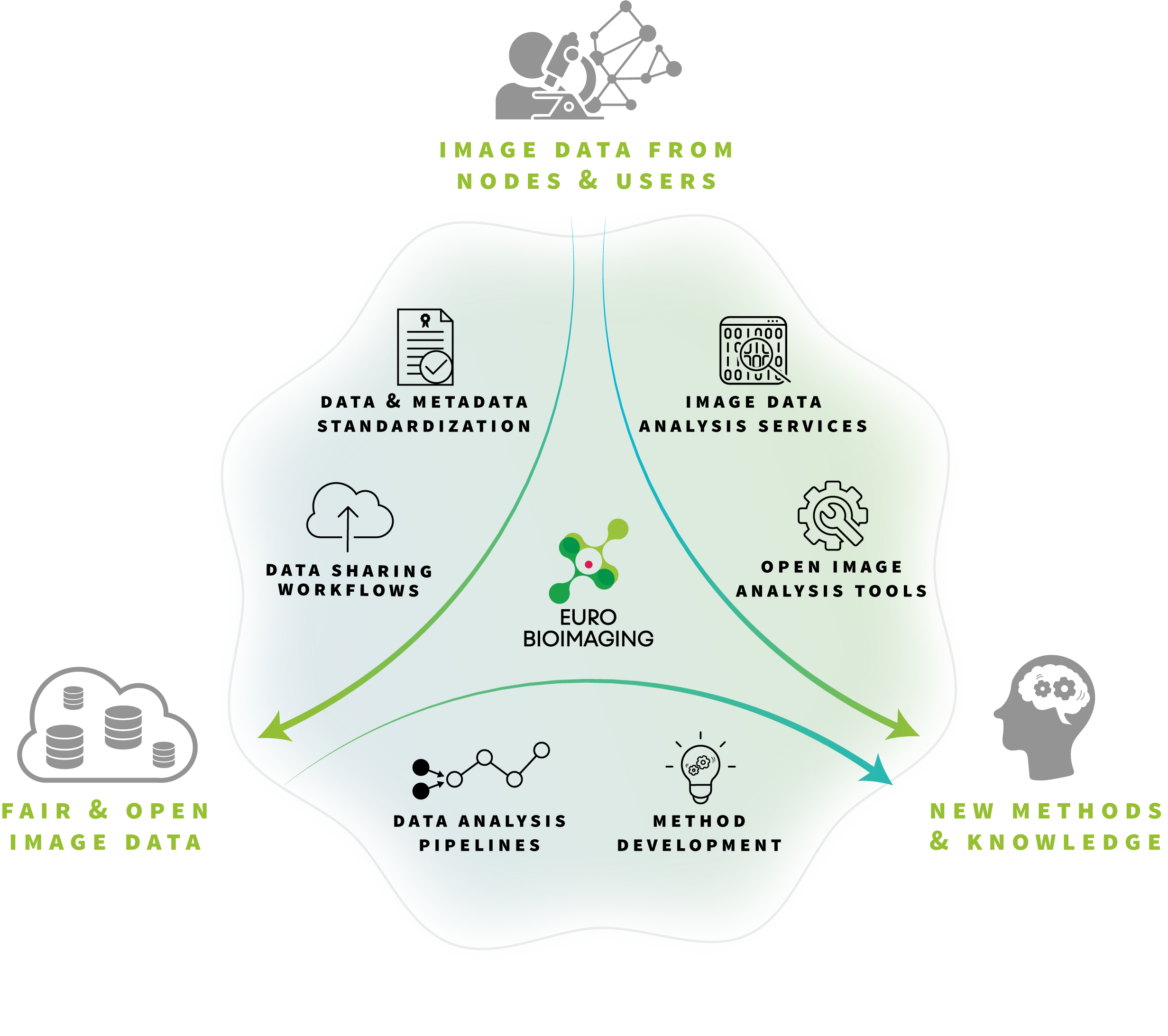
To support production of quality data, analysis methods and an extended data life cycle, Euro-BioImaging offers Image Data Services for the benefit of the whole imaging community. Together with our expert Nodes staff, we support adoption practices that yield FAIR (Findable, Accessible, Interoperable and Reusable) image data and analysis workflows.
Click on Access Services to apply for our image analysis services or click on User Data Services to learn more
Click on Access Services to apply for our image analysis services or click on User Data Services to learn more
Empowering the Community with FAIR and Open Image Data and Analysis Services