FRANCE
French BioImaging Node
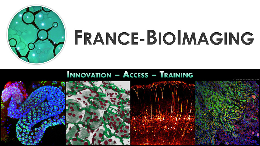
France-BioImaging (FBI) is the French national research infrastructure for biological imaging.
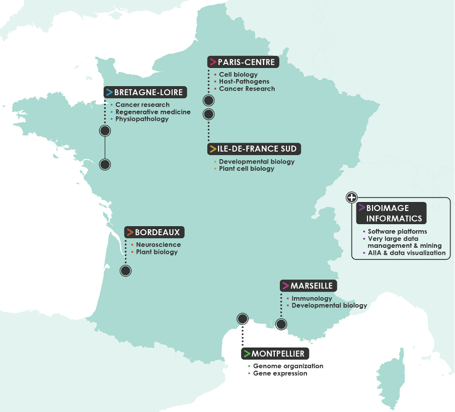
At the crossroads between molecular and cell biology, integrated physiology, biophysics and engineering, mathematics and informatics, this coordinated infrastructure gathers 23 large facilities and 60 R&D laboratories specializing in imaging in 8 regional sites (6 of which are currently open to EuBI users) and one transversal Node in bioimage informatics. FBI aims at creating the most efficient adoption of the latest advances in technologies and methods related to microscopy by the users of the imaging facilities. R&D labs agree to open their technologies and expertise to the European community either by hosting users on their own site or after technological transfer to the FBI core facilities. These technologies and methods, reinforced by a strong support in computational analysis, provide quantitative measures and integrative understanding of a wide range of cell and tissue activities in biological models, from the simplest organism, to small animals in normal and pathological situations.
Per year, we provide our ~5,000 users immediate access to cutting-edge and innovative microscopies, powerful labeling and computing methods, and appropriate training. As such, FBI plays an essential role in enabling competitive research in many fields, from fundamental questions in cell and developmental biology to preclinical research, thereby impacting a variety of domains such as agronomy, marine biology and human health. The study of human diseases is also particularly important for FBI, with many of our users working on cancer research, host-pathogen interactions, immunity, neurodegenerative and developmental disorders, genetic diseases, aging…
Offered Technologies:
ISIDORe is a Horizon Europe funded project that brings together 154 partners from 32 countries around the world, and is designed to effectively support research on infectious diseases and increase preparedness for pandemic.
| Technologies | Euro-BioImaging | ISIDORe |
| Deconvolution widefield microscopy (DWM) | ✓ | ✓ |
| Laser scanning confocal microscopy (LSCM/CLSM) | ✓ | ✓ |
| Spinning disk confocal microscopy (SDCM) | ✓ | ✓ |
| Structured illumination microscopy (SIM)* | ✓ | ✓ |
| Total internal reflection fluorescence microscopy (TIRF) | ✓ | ✓ |
| Two-photon microscopy (2P) | ✓ | ✓ |
| Objective-couple planar illumination (OCPI) | ✓ | ✓ |
| Image Scanning microscopy (ISM) | ✓ | ✓ |
| Random Illumination Microscopy (RIM)* | ✓ | ✓ |
| Lattice Lightsheet (LL) * | ✓ | ✓ |
| Single Molecula localisation microscopy (SMLM) | ✓ | ✓ |
| Stimulated emission depletion microscopy (STED) | ✓ | ✓ |
| Reversible optical fluorescence transitions (RESOLFT) | ✓ | ✓ |
| Light-sheet mesoscopic imaging (SPIM or dSLSM) | ✓ | ✓ |
| Optical projection tomography (OPT) | ✓ | ✓ |
| Macro Serial Block face Fluorecence imaging* | ✓ | ✓ |
| Raman Spectroscopy (RS) | ✓ | ✓ |
| Quantitative Phase Imaging* (QPI) | ✓ | ✓ |
| Polarization microscopy (PM) | ✓ | ✓ |
| Second/Third Harmonics Generation (SHG/THG) | ✓ | ✓ |
| High throughput microscopy/high content screening (HTM/HCS) | ✓ | ✓ |
| Fluorescence (cross)-correlation spectroscopy (FCS/FCCS) | ✓ | ✓ |
| Fluorescence Resonance Energy Transfer (FRET) | ✓ | ✓ |
| Fluorescence Recovery After Photobleaching (FRAP) | ✓ | ✓ |
| Fluorescence Lifetime Imaging Microscopy (FLIM) | ✓ | ✓ |
| Intravital Microscopy (IVM) | ✓ | ✓ |
| Voltage/pH/Ion Imaging * | ✓ | ✓ |
| Microdissection * | ✓ | ✓ |
| High-speed Imaging * | ✓ | ✓ |
| Imaging at Biosafety Level >1 | ✓ | ✓ |
| Photomanipulation | ✓ | ✓ |
| Anisotropy/Polarization Microscopy | ✓ | ✓ |
| Expansion Microscopy * | ✓ | ✓ |
| Feedback microscopy * | ✓ | ✓ |
| Multiplexing imaging *(Codex, Opal, Celldive) | ✓ | ✓ |
| Tissue Clearing (TC)* | ✓ | ✓ |
| Single molecule FRET * | ✓ | ✓ |
| TEM of chemical fixed samples (TEM) | ✓ | ✓ |
| TEM of cryo-immobilized samples (TEM cryo samples)* | ✓ | ✓ |
| Large scale EM | ✓ | ✓ |
| EM tomography (ET) | ✓ | ✓ |
| serial section TEM | ✓ | ✓ |
| Serial Blockface SEM | ✓ | ✓ |
| STEM tomography | ✓ | ✓ |
| Array tomography | ✓ | ✓ |
| Immuno-gold EM on thawed cryo-sections (Tokuyasu-EM) | ✓ | ✓ |
| Immuno-gold EM on resin sections (resin-EM) | ✓ | ✓ |
| Pre-embedding immunolabelling (pre-embed IL) | ✓ | ✓ |
| Genetic encoded EM probes | ✓ | ✓ |
| Pre-embed CLEM | ✓ | ✓ |
| on-section CLEM | ✓ | ✓ |
| Cryo Electron Tomography (Cryo-ET)* | ✓ | ✓ |
| Cryo Transmission Electron Microscopy (Cryo-TEM)* | ✓ | ✓ |
| Scanning Electron Microscopy (SEM) | ✓ | ✓ |
| pre-embeded CLEM | ✓ | ✓ |
| in-section CLEM | ✓ | ✓ |
| live-cell CLEM | ✓ | ✓ |
| CXEM (Correlative X-ray and EM) * | ✓ | ✓ |
| in vivo optical imaging | ✓ | ✓ |
| Traction Force Microscopy (TFM) * | ✓ | ✓ |
| Atomic Force Microscopy* (AFM) | ✓ | ✓ |
| Image Analysis-bio * | ✓ | ✓ |
| micro-CT | ✓ | - |
Instrument highlights
Among a number of special features offered by the France-BioImaging Node let us quote:
- Specialization in super resolution in the Bordeaux site with more than ten systems covering STED, GSD, PALM/STORM and combination of methodologies dedicated to Neurosciences and plant imaging projects
- Imaging equipment in P3 labs for host-pathogens studies in Paris sites
- Preclinical-biomedical imaging by innovative approaches in Marseille and Paris sites
Additional services offered by the Node
- Instruments
- Methodological setup (e.g. design of study protocol and standard operation procedures)
- Technical assistance to run instrument
- Probe preparation
- Animal preparation
- Animal facilities
- Wet lab space
- Data processing and analysis
- Training seminar rooms
- Housing facilities
- Regulatory affairs management service
- Biobanking, biological material storage and processing
Contact details
Edouard Bertrand (DR1 CNRS, PhD) Scientific Director of the National Research Infrastructure in Biomedical Science (INBS) France-BioImaging
Caroline Thiriet (IE CNRS)
External Affairs Manager of France-BioImaging, Node representative, EuBI user access coordinator
caroline.thiriet@france-bioimaging.org
https://france-bioimaging.org/
contact@france-bioimaging.org