NETHERLANDS
Population Imaging Flagship Node Rotterdam
The European Population Imaging Infrastructure is an initiative of the Dutch Federation of University Medical Centres (NFU) and the Department of Radiology & Nuclear Medicine, Erasmus MC, University Medical Centre Rotterdam. The ultimate aim of the infrastructure is to support the implementation of imaging in large, prospective epidemiological studies on the population and clinical level. Specific imaging biomarkers of (pre-)symptomatic diseases can be used to investigate causes of pathological alterations and to identify people at risk of developing disease or disease progression.
Specialties and expertise of the Node
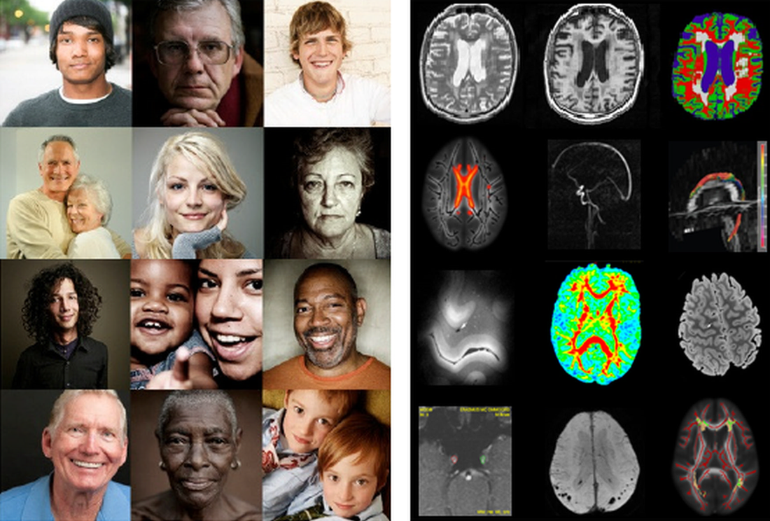
With the experience in data storage, image analysis and machine learning on large datasets acquired in the large population-based Rotterdam Study, the population imaging node aims at developing state of the art image analysis pipelines for both population and clinical studies. This Node provides technical support in and advice on 1) image storage and quality assurance 2) image analysis pipelines and 3) high volume image processing and machine learning. The population imaging flagship node collaborates with major groups that have extensive expertise in image analysis and machine learning. The node aims to offer centralized access to validated image-analysis tools and image-analysis pipelines. All pipelines are standardized, extensively tested, and metrics on the performance are available.
Offered Technologies and Services:
ISIDORe is a Horizon Europe funded project that brings together 154 partners from 32 countries around the world, and is designed to effectively support research on infectious diseases and increase preparedness for pandemic.
| Technologies | Euro-BioImaging | ISIDORe |
| Population Imaging (PI) | ✓ | ✓ |
| Image Analysis-bio * | ✓ | ✓ |
| Image Analysis-med * | ✓ | ✓ |
- Image storage facilities (XNAT based) for permanent or temporary storage of medical images
- Test-retest data image data for validation and evaluation of image analysis tools and pipelines
- Image analysis tools
- Cardiovascular disease (intracranial arterial calcification)
- Image analysis pipelines
- Neuroimaging (gray matter, white matter, cerebrospinal fluid, white matter lesions, and hippocampus)
- Cardiovascular imaging (epicardial fat, left atrial volume)
- Musculoskeletal imaging (knee cartilage, menisci)
Additional services offered by the Node
- Data processing and analysis support
- Methodological setup (e.g. design of study protocol and standard operation procedures)
- Test-retest data image data for validation and evaluation of image analysis tools and pipelines
Instrument highlights
XNAT is an open source imaging informatics platform developed by the Neuroinformatics Research Group at Washington University. It facilitates common management, productivity, and quality assurance tasks for imaging and associated data. XNAT is a hosted national service by Erasmus MC and Health-RI TraIT. It is possible to have XNAT hosted in a federated fashion, but it can also be hosted locally in your institute.
Testimonials
“We were very happy with the professional and accessible epicardial fat-analyses on our research CT-scans.” - Antonio Ribeiro, M.D., Ph.D. - ELSA-Brasil | Department of Cardiology, Universidade Federal de Minas Gerais
Contact details
Website:
www.populationimaging.eu
Stefan Klein
Department of Radiology and Nuclear Medicine
Erasmus MC, University Medical Center
Rotterdam, The Netherlands
s.klein@erasmusmc.nl
Prof. Dr. Wiro J. Niessen
Department of Radiology and Nuclear Medicine
Erasmus MC, University Medical Center
Rotterdam, The Netherlands
w.niessen@erasmusmc.nl
+31 10 70 41026
Marcel Koek
Department of Radiology and Nuclear Medicine
Erasmus MC, University Medical Center
Rotterdam, The Netherlands
i.vanhouwelingen@erasmusmc.nl