CZECH REPUBLIC
Advanced Light Microscopy and Medical Imaging Node Brno CZ
The Czech Advanced Light Microscopy and Medical Imaging Multi Modal Node in Brno provides open access to a wide range of imaging technologies and expertise to all scientists through a unified and coordinated logistics approach. The light microscopy and medical imaging units organize special programs for training scientists in biological and medical imaging techniques and data analysis.
Specialties and expertise of the Node
The Euro-BioImaging Node in Brno consists of two parts. The Medical Imaging part is formed by two closely collaborating facilities. One is focused on animal ultra-high field MR imaging and spectroscopy (9.4T), the other on human MR imaging (3T) accompanied with electrophysiological techniques. Together, these facilities enable translational research and offer a complex portfolio of MRI techniques including multimodal approaches (e.g. simultaneous EEG-fMRI) and human hyper-scanning (fMRI with two participants measured simultaneously in two scanners). The Advanced Light Microscopy part is organized as a central core facility Cellular Imaging (CELLIM) and several closely cooperating laboratories offering together access to equipment, training and image analysis tools. The light microscopy is further specialized in imaging of plant systems, mammalian germs cells, stem cells and embryos, and development of image analysis tools.
The Experimental Biophotonics Facility of this Node offers user access to their Q-Phase Multimodal Holographic Microscope developed in-house and commercialized by Tescan / Telight. The microscope provides highly quantitative and rapid measurements of cell behavior, mainly growth and motility, with unprecedented accuracy where the distribution of dry mass inside cells is determined with standard deviation of 0.4 pg/µm2. The microscopy is an implementation of Quantitative Phase Imaging where the dynamic morphometry with live cells in tissue culture is a label free technique and the accuracy is achieved by using an incoherent light source, which also uniquely allows quantitative imaging of cells in 3D environments such as collagen matrix. Integrated fluorescence imaging is available for automated time-lapse with alternating phase imaging and overlaid images are available for examination.
Offered Technologies:
| Technologies | Euro-BioImaging |
| Deconvolution widefield microscopy (DWM) | ✓ |
| Laser scanning confocal microscopy (LSCM/CLSM) | ✓ |
| Spinning disk confocal microscopy (SDCM) | ✓ |
| Structured illumination microscopy (SIM)* | ✓ |
| Total internal reflection fluorescence microscopy (TIRF) | ✓ |
| Image Scanning microscopy (ISM) | ✓ |
| Single Molecula localisation microscopy (SMLM) | ✓ |
| Light-sheet mesoscopic imaging (SPIM or dSLSM) | ✓ |
| Quantitative Phase Imaging* (QPI) | ✓ |
| Fluorescence Resonance Energy Transfer (FRET) | ✓ |
| Fluorescence Recovery After Photobleaching (FRAP) | ✓ |
| Expansion Microscopy * | ✓ |
| Tissue Clearing * | ✓ |
| micro-MRI/MRS (>= 7T) | ✓ |
| micro-MRI/MRS (>=7T) - ex-vivo | ✓ |
| Human MRI/MRS (< 7T) | ✓ |
| Image Analysis-bio * | ✓ |
| Image Analysis-med * | ✓ |
Instrument highlights
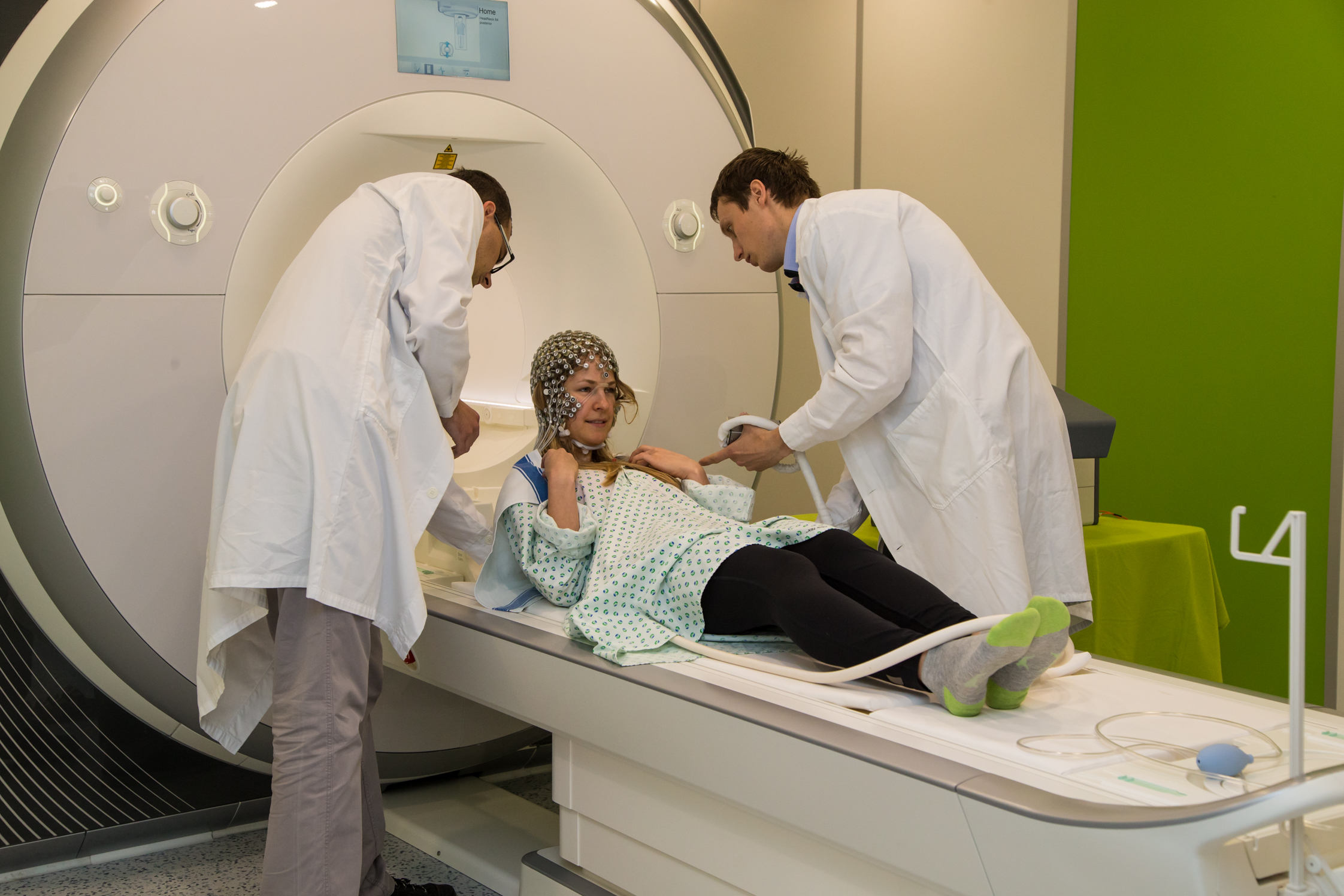
The Brno Node is equipped with two 3T Siemens Prisma scanners for human medical imaging. These scanners are designed specifically for high quality research data based on features like strong gradient fields, excellent homogeneity of mg. field and excitation, high sensitivity with 64 channel head/neck coils. Simultaneous use of two scanners enables a relatively unique feature of hyper-scanning (dual fMRI). The human MR scanners are accompanied with several MR compatible electrophysiological devices for recording of high-density EEG, ECG, breathing, skin conductance, etc.
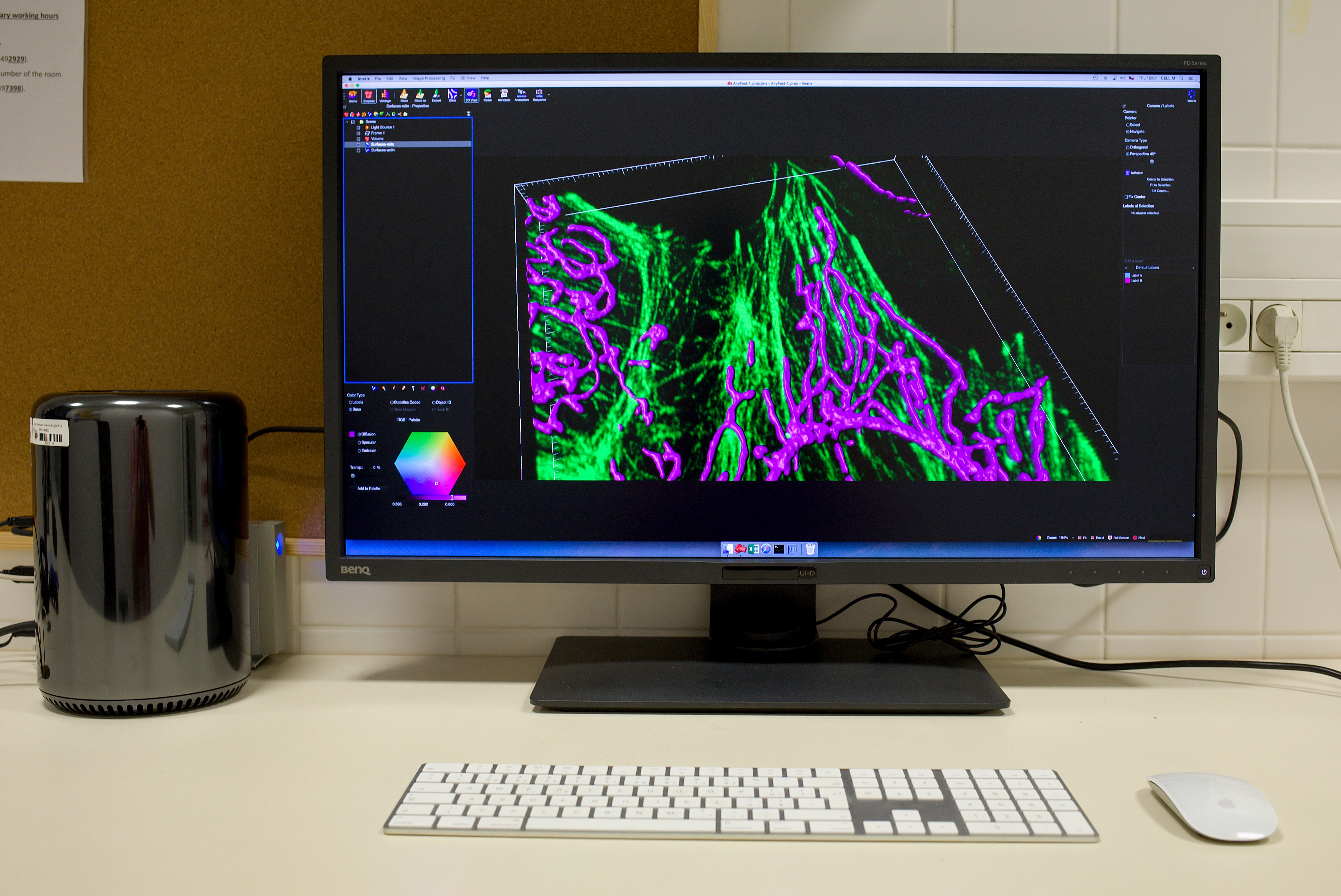
The Node also offers extensive image analysis services including tailor-made software development.
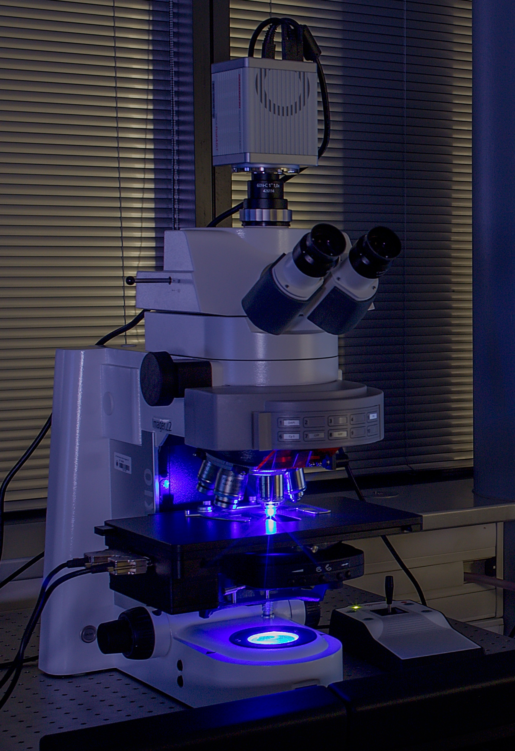
Besides providing access to a wide selection of equipment and analysis tools, the light microscopy unit specializes in plant in vivo imaging and techniques useful for research on live mammalian cells, mammalian germ cells, stem cells and embryos. The light microscopy unit CELLIM recently expanded to include a new SIM/SMLM system from Carl Zeiss, the Elyra 7 – Lattice SIM. This instrument provides several imaging modalities, like structural illumination microscopy (SIM), total internal reflection microscopy (TIRF) and single molecule localization microscopy (SMLM), which allows for a wide range of applications.
Contact details
Michal Mikl
Head of the Node
Representative of medical imaging within node
michal.mikl@ceitec.muni.cz
+00420549496099
Milan Esner
Deputy head of the Node
Representative of microscopic imaging within node
milan.esner@ceitec.muni.cz
3D view of Multimodal and Functional Imaging Laboratory:
http://www.ceitec-muni-neuroscience.pano3d.cz