AUSTRIA
Austrian BioImaging Node/CMI
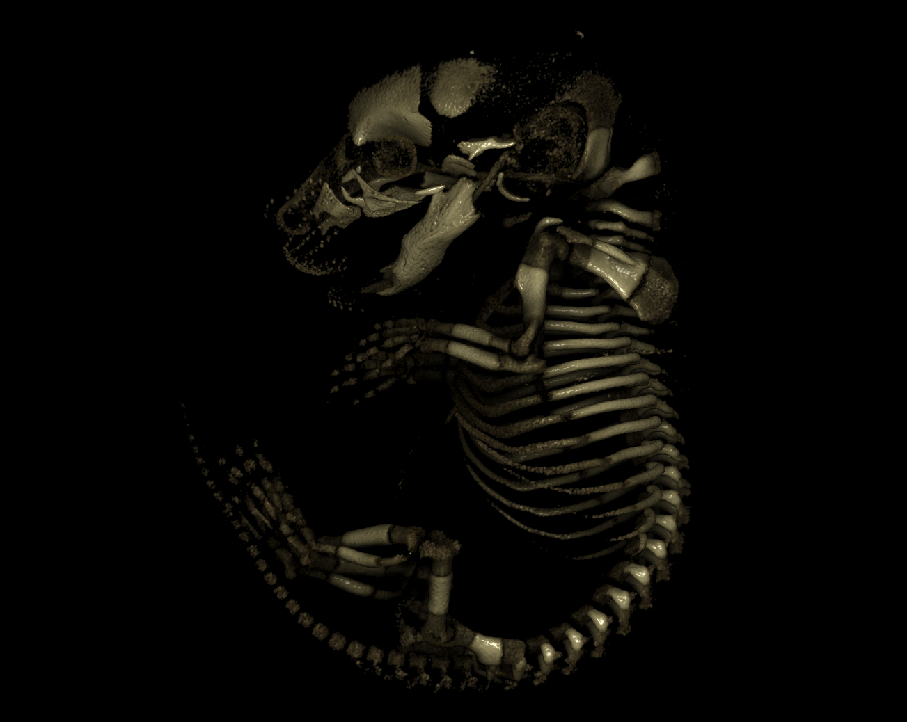
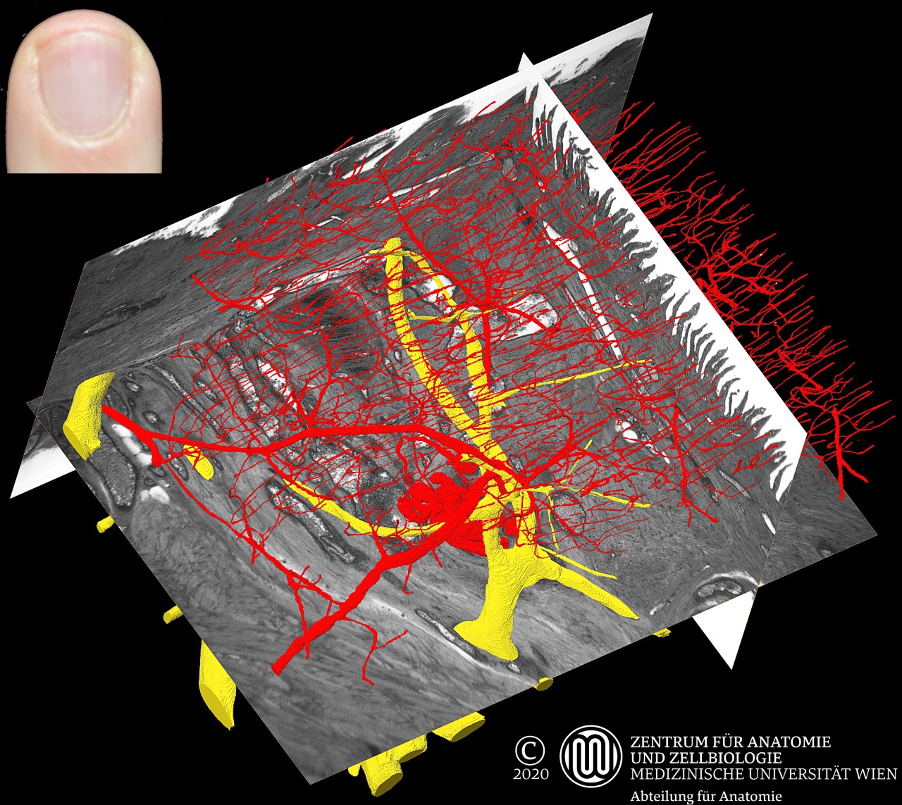
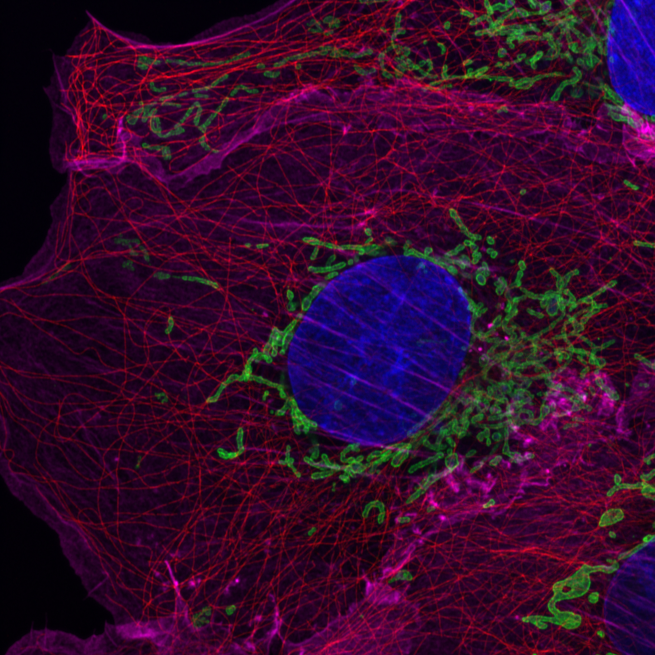
The Austrian BioImaging Node CMI (Correlated Multimodality Imaging Node) is a multi-sited, multimodality Node covering biological and biomedical imaging from cryo-electron and advanced light microscopy up to the preclinical level (including microCT, microMRI, microPET). It is hosted by 8 leading institutions across Austria with a broad service offer for organic materials, biomedical model organisms and humans, numerous multimodality imaging pipelines, and various support services, such as data and image analysis. Imaging technologies at Austrian BioImaging/CMI can be combined or used as stand-alone technologies depending on the specific biomedical research question of the user. Imaging techniques span the entire resolution range of interest for (pre)clinical and biological studies, and provide complementary sample information about structure, function, dynamics and chemical composition. More than 30 imaging techniques allow both in- and ex-vivo imaging and molecular analysis. The Node offers unique imaging techniques and expertise, such as in Optical Coherence Tomography, X-Ray Fluorescence and Mass Spectroscopy Imaging or High-Resolution Episcopic Microscopy. We are specialized in the development of advanced multimodality workflows at the forefront of correlated imaging, which can involve more than two imaging modalities.
Specialties and expertise of the Node
Austrian BioImaging/CMI has unique expertise in combining and correlating imaging technologies within multimodality pipelines to allow holistic and multiscale characterization of exactly the same sample, establishing an unprecedented and highly beneficial concept for potential customers which is solely guided by the research question at hand rather than by a fixation on a specific imaging technique. This enables Austrian BioImaging/CMI to offer individually customized imaging solutions with a variety of state-of-the-art and unique imaging technologies and support services.
Offered Technologies
ISIDORe is a Horizon Europe funded project that brings together 154 partners from 32 countries around the world, and is designed to effectively support research on infectious diseases and increase preparedness for pandemic.
Offered Technologies:
| Technologies | Euro-BioImaging | ISIDORe |
| Laser scanning confocal microscopy (LSCM/CLSM) | ✓ | – |
| Spinning disk confocal microscopy (SDCM) | ✓ | – |
| Structured illumination microscopy (SIM)* | ✓ | – |
| Total internal reflection fluorescence microscopy (TIRF) | ✓ | – |
| Two-photon microscopy (2P) | ✓ | – |
| Single Molecula localisation microscopy (SMLM) | ✓ | – |
| Stimulated emission depletion microscopy (STED) | ✓ | – |
| Photoacoustic imaging (PAI) - bio* | ✓ | – |
| Coherent Raman Anti-stokes Scattering Microscopy (CARS)* | ✓ | – |
| Raman Spectroscopy (RS) | ✓ | – |
| Fluorescence Resonance Energy Transfer (FRET) | ✓ | – |
| Fluorescence Recovery After Photobleaching (FRAP) | ✓ | – |
| Brillouin Scattering Microscopy (BSM) * | ✓ | – |
| TEM of chemical fixed samples (TEM) | ✓ | – |
| TEM of cryo-immobilized samples (TEM cryo samples)* | ✓ | – |
| EM tomography (ET) | ✓ | – |
| Focussed Ion beam SEM (FIB-SEM) | ✓ | – |
| Immuno-gold EM on thawed cryo-sections (Tokuyasu-EM) | ✓ | – |
| Immuno-gold EM on resin sections (resin-EM) | ✓ | – |
| Rre-embedding immunolabelling (pre-embed IL) | ✓ | – |
| Cryo Electron Tomography (Cryo-ET)* | ✓ | – |
| Cryo Transmission Electron Microscopy (Cryo-TEM)* | ✓ | – |
| Cryo Scanning Electron Microscopy (Cryo-SEM)* | ✓ | – |
| Cryo Focussed Ion beam (Cryo-FIB)* | ✓ | – |
| Scanning Electron Microscopy (SEM) | ✓ | – |
| micro-MRI/MRS (Field >= 7 T) (HF) | ✓ | – |
| micro-MRI/MRS (Field < 7 T)(LF) | ✓ | – |
| micro-PET | ✓ | – |
| micro-SPECT | ✓ | – |
| in-vivo micro-CT | ✓ | – |
| micro-US | ✓ | – |
| in vivo optical imaging (OI) | ✓ | – |
| PhotoAcoustic Imaging (PAI) - med* | ✓ | – |
| micro-PET/MRI | ✓ | – |
| micro-PET/CT | ✓ | – |
| micro-SPECT/CT | ✓ | – |
| Correlated Optical Coherence Tomography/PhotoAcoustic Tomography (OCT/PAT)* | ✓ | – |
| micro-MRI/MRS (>= 7T) - ex-vivo | ✓ | – |
| micro-MRI/MRS (< 7T) - ex-vivo | ✓ | – |
| micro-CT - ex-vivo | ✓ | ✓ |
| Mass spectrometry-based imaging (MSI) - med* | ✓ | – |
| Atomic Force Microscopy (AFM)* | ✓ | – |
| Micro X-ray Fluorescence Spectrometry (XRF)* | ✓ | – |
| Macro Serial Blockface Fluorescence Imaging (S-BFI)* | ✓ | ✓ |
| Image Analysis-bio * | ✓ | ✓ |
| Image Analysis-med * | ✓ | ✓ |
Additional services offered by the Node
- Project planning and methodological setup
- Wet labs
- Histology labs
- Cell culture facilities
- Animal housing
- Imaging probes
- Tracer development and radiopharmacy facility with cyclotron
- Data processing and analysis
- Data storage
Testimonials
‘Austrian BioImaging/CMI combines tremendous expertise, imaging modalities, and data management services in one organizational structure. It is managed professionally and with a lot of enthusiasm, which makes it a pleasure to work with them. I am particularly pleased with the high quality of services provided for our multimodality imaging project of mouse vasculature, the quick turnover, and also the training opportunities provided.’ Dr. Anna Obenauf, Group Leader, Research Institute of Molecular Pathology (IMP), Vienna BioCenter.
‘We were very pleased and impressed by the services provided by Austrian BioImaging/CMI for a high-quality (multimodality) analysis of our experimental samples from a a jawbone osteonecrosis project (including AFM, SEM, microCT and XRF). Many thanks for the commitment and the great work of Austrian BioImaging/CMI!’ Andrea Szabó MD, PhD, University of Szeged, Hungary.
‘Imaging support for the simulation of surgery and development of implant material at the body donor imaging unit of Austrian BioImaging/CMI was professionally supported by helpful and competent personnel. My stay was very well coordinated and I was able to produce excellent results.’ Dr. med. univ. Guan-Min Ho, Aprevent Medical Ltd.
Contact details
Baubak Bajoghli baubak.bajoghli@vbcf.ac.at
https://www.bioimaging-austria.at/