NETHERLANDS
Correlative Light Microscopy Dutch Flagship Node
The Dutch Correlative Light Electron Microscopy flagship Node is a consortium of the 4 most prominent correlative LM-EM (CLEM) labs in the Netherlands, headed by Klumperman (Utrecht), Gerritsen (Utrecht), Koster (Leiden) and Giepmans (Groningen). All labs have an international track record in microscopy technique development, application, training and open access and are at the forefront of CLEM.
Specialties and expertise of the Node
Making use of the complementary specialties of the participating labs, collectively the Node provides a unique package of CLEM methods for open access. These include state-of-the-art techniques to correlate live-cell imaging and a variety of room temperature and cryo-fluorescence microscopy techniques with immuno-EM of ultrathin cryosections, cryo-EM, 3D electron tomography and FIB-SEM. Also we can prepare samples imaged by LM in the home lab for subsequent EM analyses in our Node.
Offered Technologies:
ISIDORe is a Horizon Europe funded project that brings together 154 partners from 32 countries around the world, and is designed to effectively support research on infectious diseases and increase preparedness for pandemic.
| Technologies | Euro-BioImaging | ISIDORe |
| TEM of chemical fixed samples | ✓ | ✓ |
| TEM of cryo-immobilized samples * | ✓ | ✓ |
| Large scale EM | ✓ | ✓ |
| EM tomography | ✓ | ✓ |
| serial section TEM | ✓ | ✓ |
| Serial Blockface SEM | ✓ | ✓ |
| FIB-SEM | ✓ | ✓ |
| STEM tomography | ✓ | ✓ |
| Array tomography | ✓ | ✓ |
| Immuno-gold EM on thawed cryo-sections (Tokuyasu Method) | ✓ | ✓ |
| Immuno-gold EM on resin sections | ✓ | ✓ |
| pre-embedding immunolabelling | ✓ | ✓ |
| Genetic encoded EM probes | ✓ | ✓ |
| pre-embed CLEM | ✓ | ✓ |
| Large scale EM | ✓ | ✓ |
| on-section CLEM | ✓ | ✓ |
| CryoET * | ✓ | ✓ |
| Cryo TEM * | ✓ | ✓ |
| SEM (topography) | ✓ | ✓ |
| Elemental analysis * | ✓ | ✓ |
| pre-embeded CLEM | ✓ | ✓ |
| in-section CLEM | ✓ | ✓ |
| Cryo-CLEM * | ✓ | ✓ |
| live-cell CLEM | ✓ | ✓ |
| EDX-CLEM (Correlative EDX and SEM) * | ✓ | ✓ |
Additional services offered by the Node
- Execution of pilot studies by node staff
- Assistance with experimental design (e.g. experimental set up, choice of technique)
- Training in sample preparation
- Training in use of light and electron microscopes
- Assistance of users in the execution of experiments
- Wet lab space
- Data processing and analysis software
- Desk space and training seminar room
- Cell culture facilities
- Biological sample preparation for EM
- EM automation software
Instrument highlights
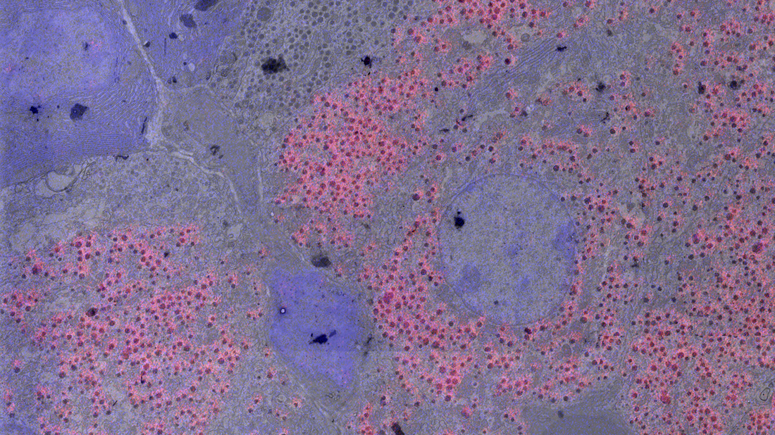
The Dutch CLEM Node contains the full set of equipment to allow CLEM experiments from light or live cell imaging to biological sample preparation to EM analysis. Specialized equipment includes cryomicrotomes, cryo-EM, iCLEM and FIB-SEM. Specific CLEM techniques include correlative light - immuno-electron microscopy on ultrathin cryosections (Tokuyasu technique), correlative live cell – 3D electron microscopy, cryo-CLEM and large volume CLEM.
Contact details
Judith Klumperman
Chair Department of Cell Biology, Head Cell Microscopy Center UMC Utrecht
j.klumperman@umcutrecht.nl
+31887556550
Additional services offered by the Node
- Execution of pilot studies by node staff
- Assistance with experimental design (e.g. experimental set up, choice of technique)
- Training in sample preparation
- Training in use of light and electron microscopes
- Assistance of users in the execution of experiments
- Wet lab space
- Data processing and analysis software
- Desk space and training seminar room
- Cell culture facilities
- Biological sample preparation for EM
- EM automation software
Instrument highlights
The Dutch CLEM Node contains the full set of equipment to allow CLEM experiments from light or live cell imaging to biological sample preparation to EM analysis. Specialized equipment includes cryomicrotomes, cryo-EM, iCLEM and FIB-SEM. Specific CLEM techniques include correlative light - immuno-electron microscopy on ultrathin cryosections (Tokuyasu technique), correlative live cell – 3D electron microscopy, cryo-CLEM and large volume CLEM.
Contact details
Judith Klumperman
Chair Department of Cell Biology, Head Cell Microscopy Center UMC Utrecht
j.klumperman@umcutrecht.nl
+31887556550