NETHERLANDS
Dutch High Field Imaging Hub
The Dutch High Field Imaging Hub is a a multi-sited, single modality Node, offering high field MRI infrastructure for human imaging. It is front runner in precision MRI focusing on medical and neuro science using advanced MRI methodologies.The five imaging centers are equipped with ultra-high Field MR instruments that can be used to obtain ultra-high resolution images of the brain, spine, breast, heart, abdomen and extremities, and benefit from increased signal to noise and new contrast associated with higher magnetic field strengths. They work closely in exchanging imaging protocols, data processing technology and RF coil technology to expedite the further development of high precision imaging in close contact with the imaging industry and national and international academic colleagues that would like to make use of the open access provided by this hub to external users.
Specialties and expertise of the Node
Whereas some of the sites excel in technical developments and translational studies in a variety of medical applications (LUMC, UMCU), other sites are more focused on more basic neurosciences oriented studies (Maastricht, Spinoza Amsterdam) with ample overlap between the sites to create a strong network that supports the knowledge base required to provide the expertise needed to support academic and non-academic user requests for ultra high field studies. This also implies that applications can be supported not only for neuro/brain studies, but for head&neck, heart, breast, pelvic and abdominal area and extremities as well.
Offered Technologies:
- Human MRI/MRS (>=7T)
Additional services offered by the Node
- Project planning and methodological setup
- Data processing and analysis
- Data storage
- Patient / Subject recruitment
- Coil lab
Instrument highlights
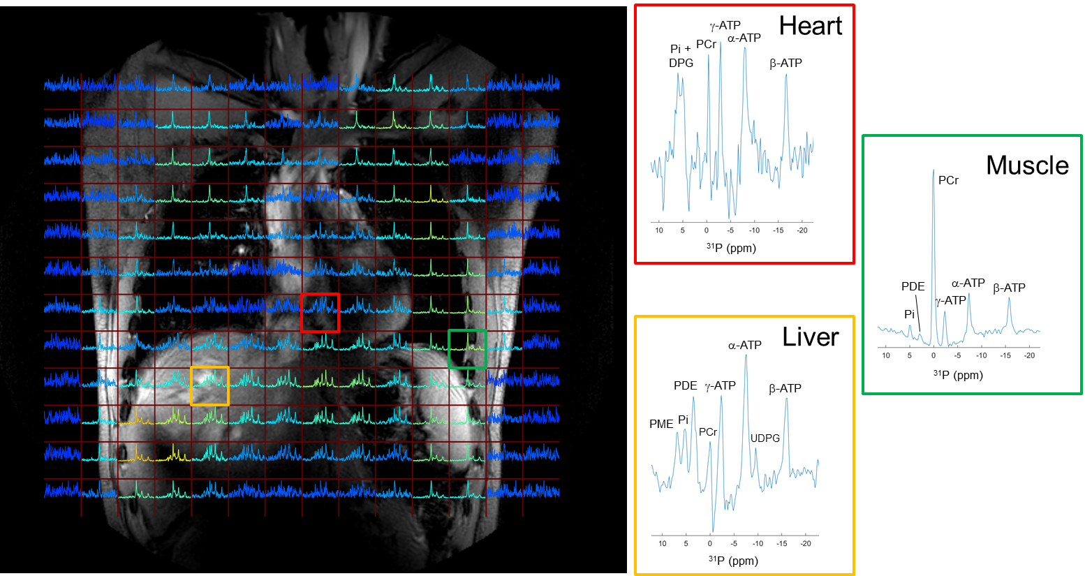
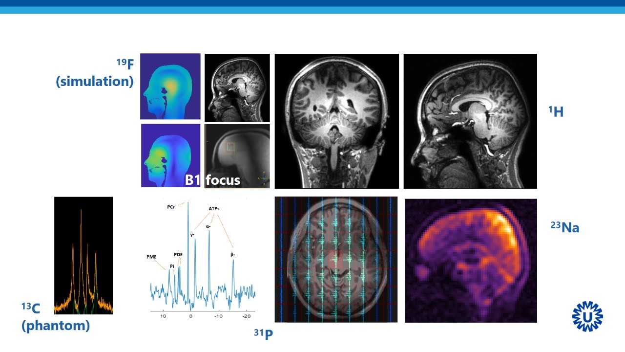
Beyond the typical 7T MRI equipment, one site of the node provides access to a 9.4T MRI scanner for human brain studies. Another site provides the capability for metabolic MRI using quintuple tuned head coil or multi tuned body coils. All sites have setups in place not only for brain studies but also for body studies.