Structured Illumination Microscopy (SIM) *
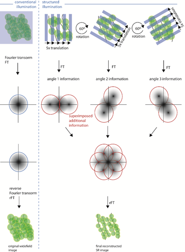
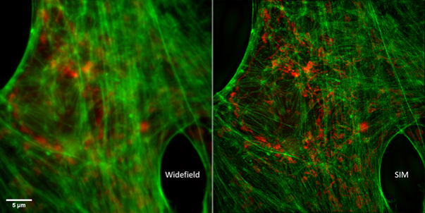
SIM belongs to the family of super-resolution microscopy techniques that allow to acquire images with higher spatial resolution than with conventional fluorescence microscopy. SIM is a camera based widefield fluorescence microscope employing a patterned illumination, e.g. stripes, to excite the fluorescence in the sample. The diffraction limited structured illumination pattern is the key element that allows to extract higher frequencies from the sample and thus achieve the improved spatial resolution. To obtain one super-resolution image, several images with shifted and rotated illumination patterns projected on the sample are acquired. The set of acquired images is then mathematically processed to reconstruct the super-resolution image. The reachable resolution improvement, both laterally and axially, is twice as better as with conventional widefield microscopy for the same wavelength. Data acquisition for one super-resolution SIM image takes from hundreds of milliseconds to seconds.
SIM is a versatile super-resolution technique used for a detailed imaging of cellular structures within cell monolayers or thin tissue sections. The strength of the method lies in the compatibility with a high number of standard fluorophores allowing relatively simple multi-color imaging, and in live-cell imaging friendliness in terms of acquisition speed and used laser powers (phototoxicity) . The downside of SIM compared to other super-resolution techniques (STED, SMLM) is a moderate resolution improvement and proneness to image artifacts formation.
There are different realizations of SIM microscopes. Euro-BioImaging nodes offer 2D-SIM, 3D-SIM and TIRF-SIM modalities on commercial setups including DeltaVision OMX, Nikon N-SIM and Carl Zeiss Elyra PS1 systems, which all use sinusoidal striped illumination.