Want to look at subcellular structures such as mitochondria, centrioles and microtubules in super-resolution? Only have access to conventional diffraction-limited microscopes? By physically expanding the sample, expansion microscopy (ExM) can achieve super-resolution images of your immunostained adherent cells. We spoke to Ana Agostinho and Steven Edwards, at the Advanced Light Microscopy facility at SciLifeLab in Sweden, part of our Swedish NMI Node. Their facility has recently added this exciting new technique to their portfolio and is now accepting applications for expansion microscopy projects as part of the Euro-BioImaging Proof-of-Concept study - along with several other Euro-BioImaging Nodes. Read more about this technology and what it can be used for in the interview below.
We are today talking about Expansion Microscopy imaging. Please provide a short summary of this type of imaging and list some applications:
Ana: My name is Ana Agostinho and I am a staff scientist at the Advanced Light Microscopy facility at SciLifeLab in Stockholm, Sweden which is part of the Swedish National Microscopy Infrastructure.
Steve: My name is Steven Edwards and I am working together with Ana at the Advanced Light Microscopy facility at SciLifeLab, Stockholm.
Ana: Our facility has been providing scientists with advanced sample preparation support for super-resolution microscopy techniques such as STED and SMLM. We have recently started offering scientists support with expansion microscopy, which enables super-resolution microscopy to be performed on diffraction limited microscopes. Without going into too much detail, the method involves crosslinking proteins in a fixed sample to a swellable polymer, which can be physically expanded by immersion in distilled water.
Steve: The technique is rapidly developing with many new articles and protocols published each year. We realised that this could be somewhat daunting for a biologist wishing to start with the method. We have chosen a single protocol, based on Magnified Analysis of the Proteome which is robust in our hands. It allows modest expansion (4-5x) of adherent cells.
Ana: We provide scientists with the reagents for gelation and digestion as well as access to simple laboratory equipment such as an oven and Eppendorf heater/shaker. We also give suggestions for sample mounting which must be stable enough to allow for long microscope acquisitions without drift.
Tell us a bit more about a specific project that was done in your facility using this technology? What scientific questions were you addressing?
Steve: Much of the work to-date has been focused on validating different expansion protocols and assessing their suitability for use in a facility environment. In order to provide adequate support, we have been focusing on the expansion of immunostained adherent cells.
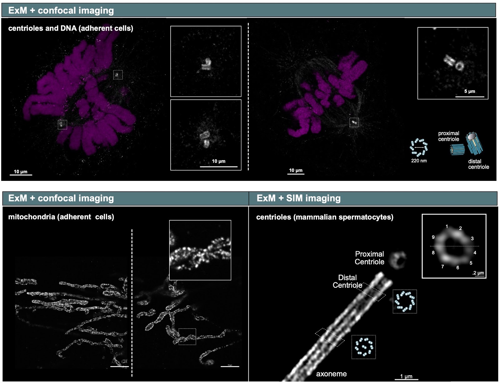
Ana: With that being said, using this technique, in combination with confocal imaging, we have been able to clearly visualise the general architecture of mammalian centrioles (200 nm in diameter), as well as the fine details of the mitochondria inner membrane, typically imaged with super-resolution techniques (e.g. STED) (Figure, top panel and lower left panel). In addition, we have tested the possibility of further increasing the resolution when imaging our gels by combining expansion microscopy with super-resolution imaging modalities such as SIM and lattice SIM. This has enabled us to visualise the 9 microtubule triplets that form the centriole and fine details of the axoneme, in mammalian spermatocytes (Figure, lower right panel).
Example of Expansion Microscopy compared to other approaches (see text above for explanation). Image courtesy of Ana Agostinho, SciLifeLab, Swedish NMI.
What are some advantages of this technique that make it suited to addressing these types of questions?
Ana: Expansion microscopy is versatile with regards to the antibodies you can use to detect proteins of interest. In this case, we could visualise sub-cellular structures using the same primary antibodies we had already established in the lab, achieving sub 200 nm resolution on a confocal microscope.
Steve: The advantages of expanding samples are not limited to the spatial resolution improvement. Staining may also be improved as proteins are de-crowded and additional epitopes are made available.
What other services do you provide in your facility that would be useful in combination with this type of imaging?
Steve: We are fortunate to have a wide array of microscopes available to image expanded samples. Sub-50 nm resolution can be achieved with a simple point scanning confocal, but the airyscan detector from Zeiss is particularly suitable for detecting the weak signal from expanded samples. Successful application of this technique requires that the samples are treated with the same “care” that is required for other super-resolution modalites. We can therefore provide scientists with advice on fixation and labelling as part of the service.
How to apply:
Expansion Microscopy is part of the Euro-BioImaging Proof-of-Concept study - and it's available at multiple Nodes. The Proof-of-Concept study makes it possible to introduce exciting, new imaging technologies to our portfolio that were previously unavailable via our network. We are currently accepting applications to use these technologies at participating Nodes as part of the Proof-of-Concept study. Be part of this study - and contribute to community-wide continuous technological innovation!
All scientists, regardless of their affiliation, area of expertise or field of activity can benefit from Euro-BioImaging’s pan-European open access services. Potential users of these new technologies are encouraged to submit project proposals via our website. To do so, you can log in to access our application platform, choose the technology you want to use and the facility you wish to visit, then submit your proposal. All applications will be processed by the Euro-BioImaging Hub. As usual, users will benefit from advice and guidance by technical experts working at the Nodes, training opportunities, and data management services.
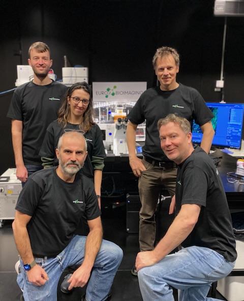
ALM facility at SciLifeLab, Stockholm: Top left: Steven Edwards, Top middle: Ana Agostinho, Top Right: Stefan Wennmalm, Bottom Left: Hjalmar Brismar, Bottom Right: Hans Blom
More news from Euro-BioImaging
July 15, 2026
Imaging-Driven Neuroscience, Responsible Research, and the Power of Advocacy: Highlights from FENS Forum 2026
Euro-BioImaging was pleased to participate in the FENS Forum 2026, Europe’s largest international gathering dedicated to advancing neuroscience (>8000 participants). Throughout…
Euro-BioImaging Highlights the Value of Research Infrastructures at the FENS 2026 Satellite Symposium hosted by EBRAINS
Euro-BioImaging was pleased to participate #FENS2026 Satellite Symposium “Accelerating Your Neuroscience Research through the European Research Infrastructures” organised by EBRAINS, to learn…