
October 16, 2026
RAISE Hub: One gateway to Europe’s AI in Science Community
A new online platform to connect researchers, infrastructures, and funders to accelerate AI scientific discovery across Europe Dublin, Ireland, 15 October…



Many Euro-BioImaging Nodes are working on innovative image analysis and image data management solutions and the next Euro-BioImaging User Forum is designed to highlight these. Join us for an afternoon of presentations from Euro-BioImaging users and image data/image analysis experts at our Nodes that will provide a compelling overview of the state-of-the-art in Image Data. At this event, Anne-Sophie Mace, Institut Curie, part of France-BioImaging Node, will explain how the FBIAS, France BioImage Analysts, provides customised bioimage analysis advice & workflows through a nationwide, remotely operating core-facility
What: Euro-BioImaging User Forum “Image Data”
When: March 26, 2024, from 14:00-17:00 CEST Where: Online
Abstract
Providing customised bioimage analysis advice & workflows through a nationwide, remotely operating core-facility
Anne-Sophie Mace
Institut Curie, FBI Node
FBIAS, France BioImage Analysts, was created in 2021 with the ambitious objective to build a nation-wide remotely operating core facility for bioimage analysis service.
The first step consisted in creating a network of bioimage analysts located throughout France and organising (remote) monthly meeting to discuss technical issues linked to users’ projects. In 2022, FBIAS started offering advice on image analysis to the biologists of the France BioImaging (FBI) national infrastructure, in the form of open desks. Every 2 months, users from all over the France can book a 1-hour slot with 2 or 3 bio-image analysts to discuss their project. The purpose is to support the users either by solving a specific problem or by advising them on the best practices and tools to use, within the 1-hour slot.
However, 1 hour is often not sufficient because users need customised workflows to answer a problematic very specific to their research. To illustrate this, we will present a project developed within the Curie Institute which aims at finding back in a fixed sample cells that were first imaged and tracked in a 48-hours movie. The implemented strategy is based on registration of the contours of the biological structures, acquired in different modalities, performed at the sample scale. The designed workflow offers flexibility with easy intervention of the user at each step of the process, necessary to treat correctly all the data (even distorted samples). It was successfully used on wide and thick samples (organoids, brains) to follow cell development whose fate has been determined thanks to the immunostaining.
To make this remote core facility really operational, answering demands of biologists all over the France and maybe outside, some challenges are still to achieve: a common charging system, a committee evaluating the projects and of course more engineers to create the customised workflows.

October 16, 2026
A new online platform to connect researchers, infrastructures, and funders to accelerate AI scientific discovery across Europe Dublin, Ireland, 15 October…
October 16, 2026
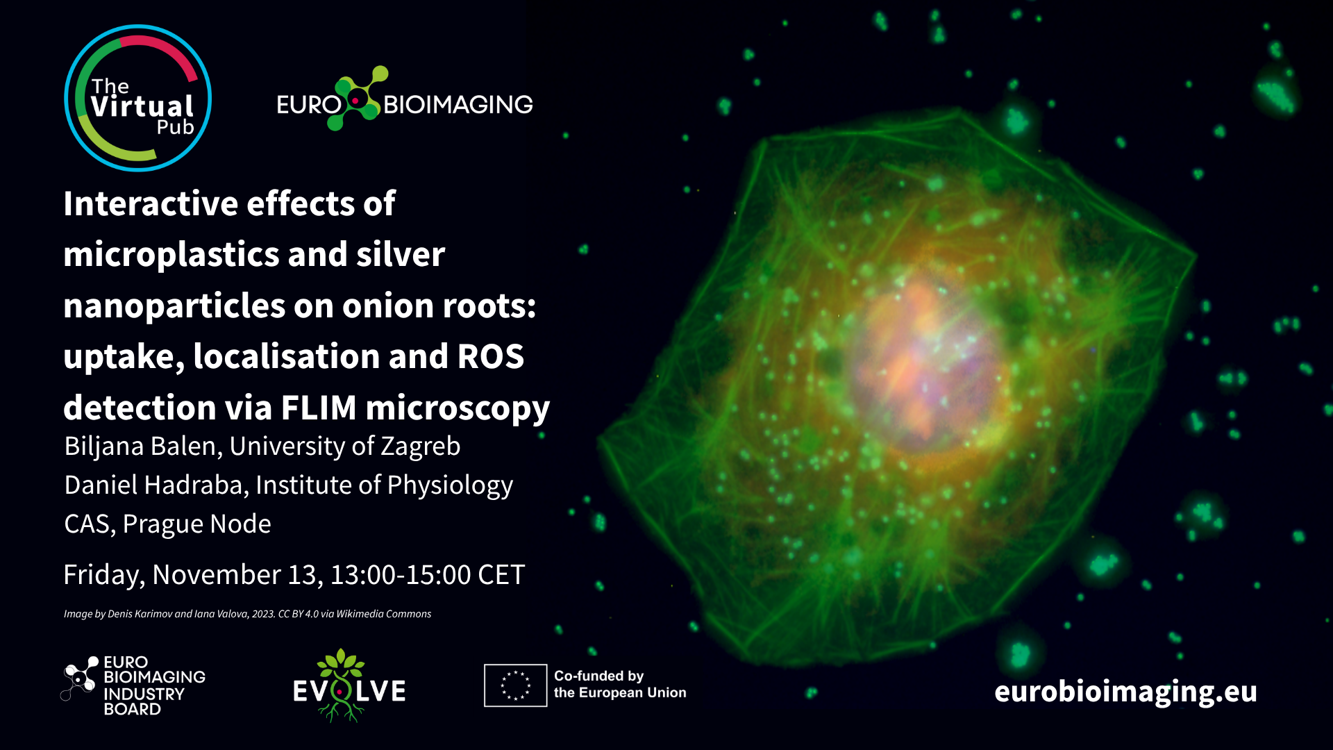
At the Special Edition Virtual Pub, “Imaging Micro- and Nanoplastics,” Euro-BioImaging users, Node experts and Industry partners will explore how advanced microscopy, clinical…
October 16, 2026
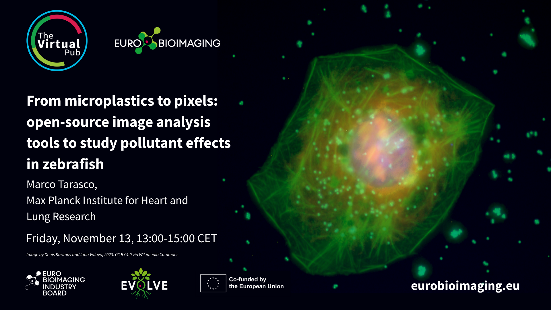
At the Special Edition Virtual Pub, “Imaging Micro- and Nanoplastics,” Euro-BioImaging users, Node experts and Industry partners will explore how advanced microscopy, clinical…