Euro-BioImaging is organizing an online User Forum on Tuesday, March 21, 2023, from 14:00-17:00 CET. The topic is “Cardiovascular research.” This event will highlight the importance of cutting-edge imaging technologies in support of cardiovascular health, disease, diagnostics and the development of therapies. We will showcase the specific expertise available at our Nodes across Europe through case studies presented in tandem with the research community.
At this event, Eva Rog-Zielinska, University of Freiburg, will explore how dual axis electron tomography, performed in collaboration with the EMBL Node, allows for the investigation of 3D nanostructure at different stages of the contraction–relaxation cycle of individual cardiomyocytes.
Cardiac muscle cells, cardiomyocytes, undergo significant deformation during every heartbeat. The contraction of the heart alters cellular ultrastructure, which in turn modulates cell function. The exact interplay between deformation, ultrastructural dynamics, and function is hard to predict from macro- or micro-scale assays. As it is experimentally challenging to measure nanoscopic dynamics, the effects of cardiomyocyte deformation on the intracellular ultrastructure (e.g. organelle shape or position) are largely unexplored. Action potential-synchronised high-pressure freezing of cardiomyocytes, coupled with dual axis electron tomography, allows for the investigation of 3D nanostructure at different stages of the contraction–relaxation cycle of individual cardiomyocytes. We present the experimental workflow and the initial quantification of the organelle deformation during rest and peak contraction of cardiomyocytes. We plan to fuse this information into a statistical model of nanoscopic cardiomyocyte contraction dynamics, which will help us understand how ultrastructure–function relationships contribute to cellular steady-state and its pathological alterations in disease.
More news from Euro-BioImaging
May 29, 2026
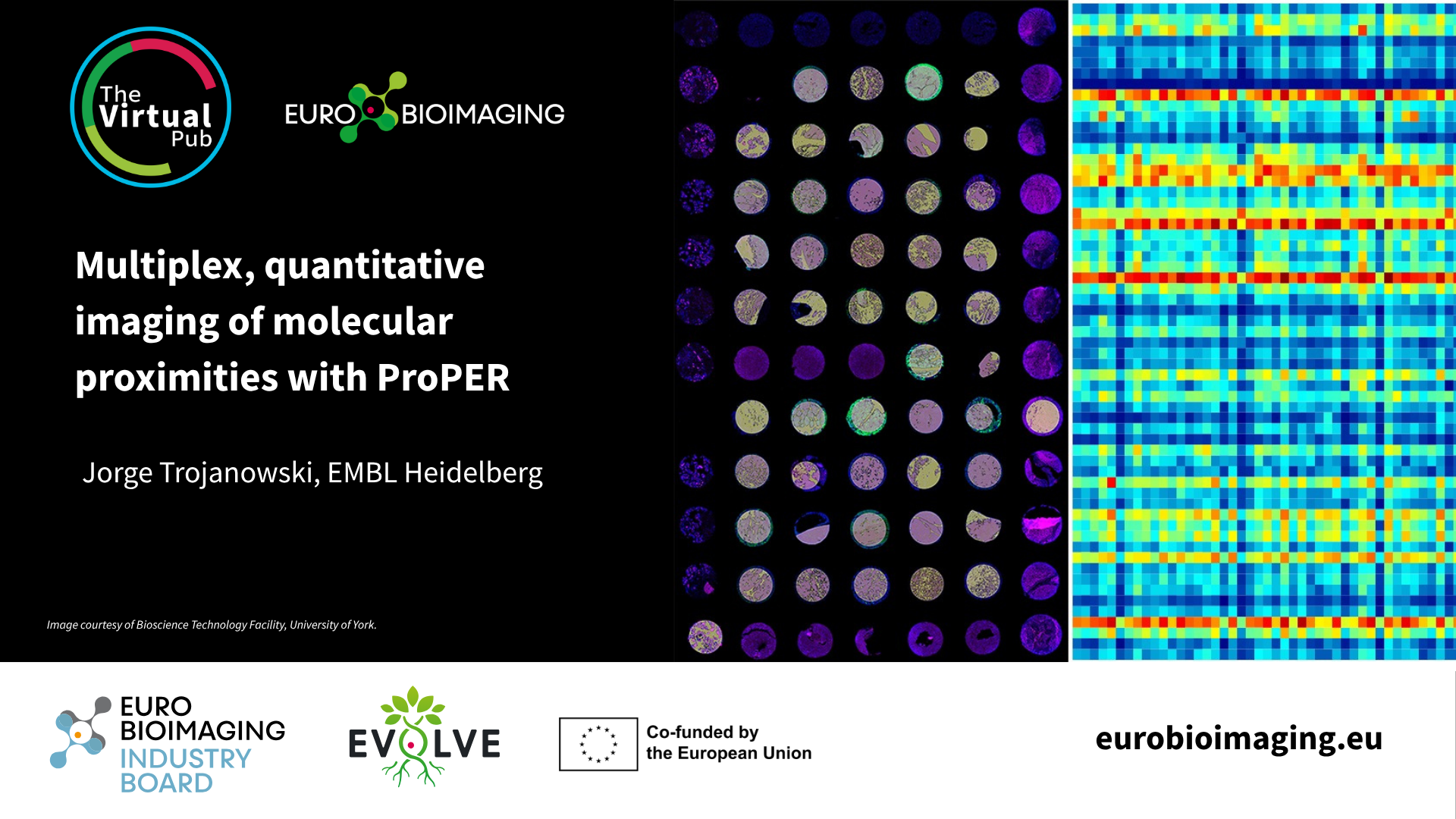
Multiplex, quantitative imaging of molecular proximities with ProPER
Spatial transcriptomics is rapidly transforming the way researchers study complex biological systems. To explore the latest developments in this fast-moving field, Euro-BioImaging is…
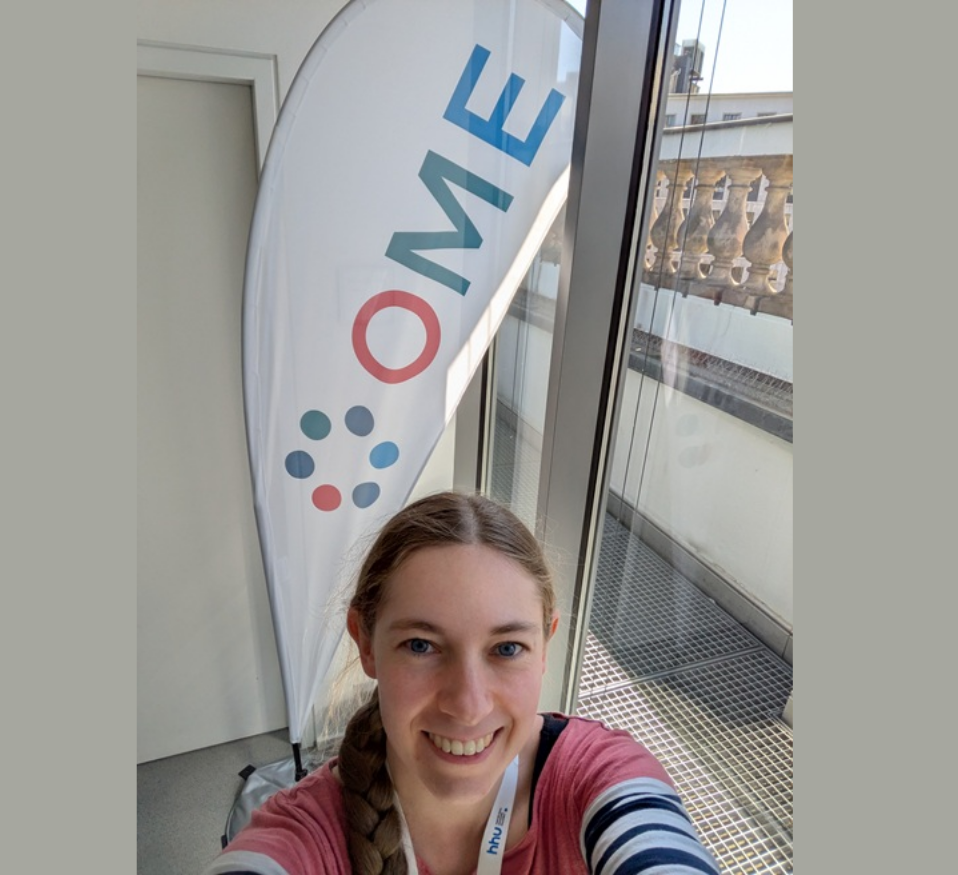
Euro-BioImaging at OME26: Shaping the Future of Open Imaging Data
The OME2026 Community Meeting, held from 28.04.2026 to 30.04.2026 in Düsseldorf and online, brought together developers, imaging scientists and data experts from across…
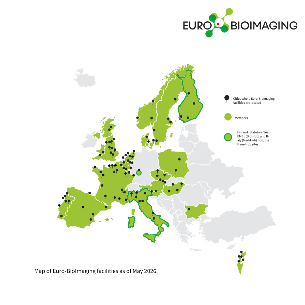
Euro-BioImaging is delighted to announce a significant expansion. Six of our Nodes – Austrian BioImaging/CMI, UK Node, Cellular Imaging Hungary, Danish BioImaging, Portuguese…