The origin and evolution of insular large mammals is key to understanding broader mammalian adaptations and selective processes, offering valuable insights relevant to other insular species, including extinct hominins and extant species. Dr. Luca Pandolfi, from the Department of Earth Sciences of the University of Pisa, is specialized in vertebrate paleontology and paleobiology, with a focus on mammalian evolution and adaptation. He accessed the IFC CNR facility of the MultiModal Molecular Imaging Italian Node with SEELIFE funding to perform CT scanning and 3D reconstruction of fossil skulls, getting insights into the evolution of these mammals and their adaptation to ecological niches.
For decades, studies using ancient biomolecules and classical morphometrics have aimed to reconstruct the evolution of extinct Eurasian mammals. However, conclusive evidence regarding the origin of many species inhabiting Mediterranean islands during the Pleistocene remains elusive. Dr. Luca Pandolfi investigates the origin and evolution of large mammals, such as Cervidae, Hippopotamidae and Elephantidae, from the central Mediterranean islands (e.g., Sicily, Malta). In particular, he is interested in the brain's morphology in extinct species, which provides critical insights into their potential cognitive capabilities, sensory adaptations, and even aspects of social behavior and locomotion. “Reconstructing brain evolution offers a unique window into how lineages adapted to past environments and ecological niches”, he says.
Working with fossil skulls
A central part of this research involves combining spatial distribution modeling with detailed 3D morphometric analyses of internal and external cranial structures. This can be achieved through Computed Tomography (CT) scanning, a powerful, non-destructive and cost-effective technique suitable for studying large sample sets. CT scanning and 3D reconstruction of the endocranial cavity within fossil skulls of hippopotami and cervids from Africa and Mediterranean Basin, including islands, provided by the Natural History Museum of the University of Pisa, have been conducted at the state-of-the-art facilities of the CNR Institute of Clinical Physiology (IFC) in Pisa, part of our Multi Modal Molecular Imaging Italian Node.
“Moving and positioning the heavy and cumbersome skulls was not an easy task, and careful instrument calibration was needed to adapt to the skull mineral composition, which is highly variable across different fossil samples. The facility staff at IFC was very supportive and professional.”
This study project builds on Dr. Pandolfi’s earlier work, also carried out at IFC, which focused on high resolution micro-CT scanning of the enamel-dentine junction in suids from the hominoid-bearing localities of the Tusco-Sardinian archipelago. Taken together, the two projects define a coherent and innovative research strategy in which advanced imaging is used to extract hidden anatomical information from fossil remains, combining dental, cranial, and endocranial evidence to reconstruct taxonomy, evolutionary relationships, and adaptive change in insular vertebrates.
Generating 3D datasets
Acquiring the CT scans on the fossil skulls required a huge amount of preliminary work. “Moving and positioning the heavy and hindering skulls was not an easy task, and careful instrument calibration was needed to adapt to the skull mineral composition, which is highly variable across different fossil samples. The facility staff at IFC was very supportive and professional”, Dr. Pandolfi says.
The CT scans have already generated high-resolution three-dimensional datasets of the fossil crania, providing non-destructive access to internal anatomical structures that are otherwise difficult to investigate in rare and fragile specimens. This represents a substantial achievement of the project, because it makes it possible to document cranial architecture in detail and to establish a robust digital basis for subsequent analyses. In particular, these datasets will support the reconstruction of the endocranial cavity and the reconstruction of virtual endocasts, which are essential for investigating variation in brain size and shape in extinct insular mammals.
Image analysis
At present, some interpretations are already ongoing, but digital segmentation and the full quantitative morphometric analyses are still in progress. Once completed, these analyses are expected to allow detailed comparisons between insular dwarf species and their possible mainland relatives, helping to clarify the extent and direction of morphological divergence associated with long-term isolation in island environments. This will be particularly relevant for evaluating broader evolutionary questions such as the phylogenetic relationships of the studied taxa and the extent to which their anatomical changes may be consistent with patterns commonly discussed under the “island rule.” In addition, the endocranial data are expected to provide new paleoneurological information, including evidence on relative changes in brain proportions and on sensory adaptations that may reflect different ecological pressures in insular settings.
Important insights into evolution in island settings
More broadly, this research is expected to provide a useful comparative framework for understanding the evolution of other insular vertebrates, both extinct and extant. Examples include extinct hominins such as Homo floresiensis and H. luzonensis, whose insular histories have raised important questions about the relationship between body size, brain evolution, and ecological adaptation, as well as living island mammals such as Sus philippensis and Cervus elaphus corsicanus. In this sense, the present study is not limited to the Mediterranean fossil record, but contributes to a wider discussion on how isolation, resource limitation, and ecological specialisation shape cranial and neuroanatomical evolution across island systems.
“Access to the MMMI Node and getting financial support from SEELIFE was a key step for this project and in my research trajectory. It provided an important opportunity to address a relevant question in evolutionary paleontology, to apply and further develop advanced methodological approaches, and to strengthen my research profile at the University of Pisa.”
- Dr. Luca Pandolfi, University of Pisa
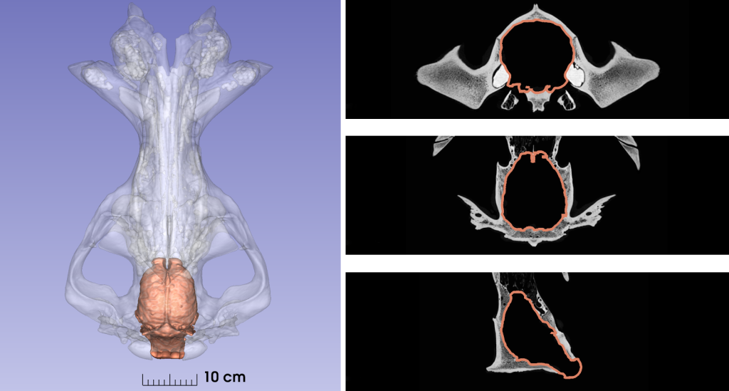
A representative hippopotamus (Hippopotamus amphibius) skull CT scan acquired on a Siemens Biograph mCT64 scanner, with endocranial segmentation for morphological quantitative measurements.
More news from Euro-BioImaging
July 27, 2026
Moara Lemos: New insights from Global BioImaging’s Job Shadowing programme
Moara Lemos is a Brazilian researcher and imaging scientist. Her award winning research pushes the boundaries of structural biology, using advanced cryo-electron microscopy…
A Multiplexed Vision: Highlights from the Special Edition Virtual Pub on Spatial Transcriptomics
On June 12th, Euro-BioImaging hosted its latest Special Edition Virtual Pub, bringing together over 200 researchers and technology developers working at the cutting…
Cellular Imaging Hungary Node expands expertise in advanced neurophotonics
The Euro-BioImaging Cellular Imaging Hungary Node has expanded its service portfolio with the addition of the BrainVisionCenter (BVC) in Budapest. Following a successful…