

July 9, 2026
Euro-BioImaging Visits the NORMOLIM Node at the 2026 180°N Conference
In April 2026, the Med-Hub Head of Operations Alessandra Viale attended the 2026 180°N Conference, held at the beautiful Nye Hjorten Teater…
Q: Please tell us who you are, where your facility is based, and what your role is.
Antti: My name is Antti Isomäki and I am an optical physicist by training. The path to my current position went through the basic research in a laser lab, postdoc time in microscopy technology development, and finally biomedical applications and imaging method development. I have been working at the Biomedicum Imaging Unit for more than eight years now and, as my main role, I provide support for the label-free nonlinear imaging techniques. Our core facility is located at the medical campus of the University of Helsinki.
Q: We are today talking about CARS Microscopy. Please provide a short summary of this type of microscopy and list some applications.
Antti: Coherent Anti-Stokes Raman Scattering microscopy (CARS) offers complimentary contrast in samples where we do not want or cannot use fluorescent labels. As the technique is based on resonantly driven intrinsic molecular vibrations, the strength of the observed signal depends heavily on the molecule concentration in the focus.
In the beginning, our main applications were related to imaging lipid droplets in fixed and live cells. Another promising application has been imaging unstained tissue sections, taken from e.g. liver biopsies, where CARS signal from lipids and second harmonic generation from collagen fibers can be detected simultaneously.
More recently, CARS has proved to be extremely powerful in imaging pharmaceutical materials. We have demonstrated high-resolution chemically specific imaging of drug microparticles in cell cultures and tissue. In pharmaceutical material development, CARS can distinguish between various non-fluorescent materials, or even between the polymorphs of the same drug material. This information is very useful when, for example, tablets with optimal dissolution profiles are developed.

Q: Tell us a bit more about a specific project that was done in your facility using this technology? What scientific questions were you addressing?
Antti: Researchers from the field of pharmaceutical chemistry and technology have combined CARS with various other methods to analyze drug crystallization and its influence in dissolution of tablets. Crystallization during storage and administration is a major problem that affects the efficacy of the drug. Using CARS it was possible to detect subtle amounts of crystals, which may be important when developing new drugs and dosage forms. Here is an example of how CARS can be used to understand dissolution and crystallization:
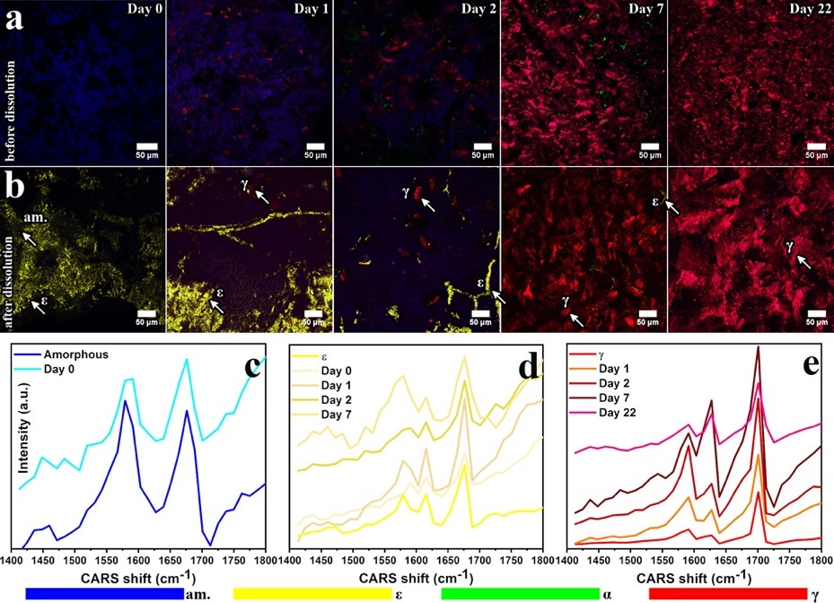
CARS and SFG overlay images of samples stored at 30 °C/23% RH before (a) and after (b) 15 min of dissolution testing at pH 6.8. CARS spectra in the range of 1413–1800 cm-1 from selected regions marked by arrows plotted with reference spectra of amorphous, ε-, and γ-indomethacin (c–e). Overlay images (a, b) represent overlays of three channels: single CARS line at 1701 cm-1 in red (γ-indomethacin), single CARS line at 1676 cm-1 in blue (amorphous indomethacin), and a third channel representing the SFG signal (all noncentrosymmetric crystals) in green and yellow. The separation between green and yellow regions is based on the intensity ratios of CARS peaks at 1652 and 1676 cm-1 so that the regions having SFG activity and a CARS peak at 1652 cm-1 are colored green (α-indomethacin), and the regions having SFG activity and a CARS peak at 1676 cm-1 are colored yellow (ε-indomethacin). Panel (a) has been reprinted with permission from Novakovic et al.(18) Copyright 2017 American Chemical Society. am = amorphous. (Source: Novakovic et al., Understanding Dissolution and Crystallization with Imaging: A Surface Point of View, Mol. Pharmaceutics 2018, 15, 11, 5361–5373 https://doi.org/10.1021/acs.molpharmaceut.8b00840) Further permission related to the material should be directed to the ACS.
Q: Why is this technology best suited to address that question?
Antti: CARS together with other nonlinear microscopy modalities allows for fast, high-resolution imaging of very early stage crystallization that was not detectable with the conventional solid-state analysis methods such as X-ray powder diffraction (XRD) or Attenuated total reflectance – Fourier transform infrared spectroscopy (ATR-FTIR).
Q: What are some challenges of this kind of microscopy? What do researchers have to pay attention to when performing these experiments?
Antti: Often the sample preparation is easier than with fluorescence-based methods – you simply have your unstained cell culture, tissue or other sample on a cover slip. Image scanning works largely the same as in confocal microscopy. However, the interpretation of the resulting images requires some careful consideration due to the nonlinear nature of the method. For example, quantitative measurements are challenging. CARS is also not as sensitive as fluorescence imaging, so one should not expect single molecule precision.
Q: What other services do you provide in your facility that would be useful in combination with this type of microscopy?
Antti: The imaging facilities in the Helsinki Node provide access to most of the commonly used microscopy techniques varying from in vivo optical microscopy to electron microscopy. Therefore, multimodal imaging combining CARS with other techniques is possible here. As an interesting example, we have demonstrated correlative CARS and electron microscopy to study cell-nanoparticle interactions [1]. We collaborate also with other facilities and departments, and can help getting access to additional sample preparation and analysis methods.
[1] Saarinen, J., Gütter, F., Lindman, M., Agopov, M., Fraser‐Miller, S.J., Scherließ, R., Jokitalo, E., Santos, H.A., Peltonen, L., Isomäki, A. and Strachan, C.J. (2019), Cell‐Nanoparticle Interactions at (Sub)–Nanometer Resolution Analyzed by Electron Microscopy and Correlative Coherent Anti‐Stokes Raman Scattering. Biotechnol. J., 14: 1800413. doi:10.1002/biot.201800413
Q: Why should scientists choose your facility for using this technology?
Antti: We were among the first sites to start working with the commercial CARS microscope from Leica. During the years, we have tested quite a large number of different samples and got a good understanding of the possibilities and limitations of the technique. I am always ready to discuss the feasibility or run a pilot test to see if our instrument works for certain application. Finally, I should mention that some new instrumentation for CARS and stimulated Raman spectroscopy (SRS) will be available in Helsinki next year. I hope that by that time the scientists will be able to travel here more freely again.
In July 2020, Euro-BioImaging launched a Proof-of-Concept study - in collaboration with its Nodes - making it possible to access six previously unavailable imaging technologies. This article – the fourth in a series - features interviews with Euro-BioImaging Node staff, shedding light on some potential applications of these technologies - and providing advice on how to get the most out of them. We are currently accepting applications to use these technologies as part of the Proof-of-Concept study.
All scientists, regardless of their affiliation, area of expertise or field of activity can benefit from Euro-BioImaging’s pan-European open access services. Potential users of these new technologies are encouraged to submit project proposals via our website. To do so, you can log in to access our application platform, choose the technology you want to use and the facility you wish to visit, then submit your proposal. All applications will be processed by the Euro-BioImaging Hub. As usual, users will benefit from advice and guidance by technical experts working at the Nodes, training opportunities, and data management services.
For more information: info@eurobioimaging.eu
July 9, 2026
In April 2026, the Med-Hub Head of Operations Alessandra Viale attended the 2026 180°N Conference, held at the beautiful Nye Hjorten Teater…
July 7, 2026
Armed conflicts generate long-lasting environmental contamination that extends well beyond the duration of military operations. The release of heavy metals such as Arsenic,…
July 6, 2026
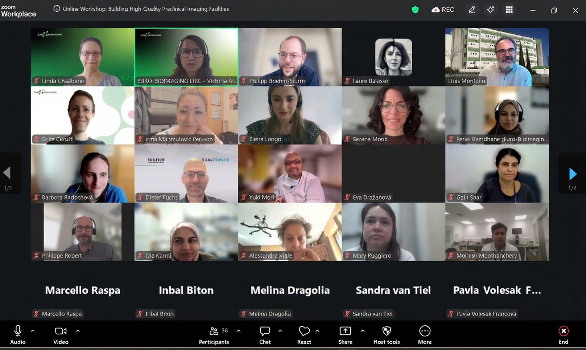
On 2 July 2026, Euro-BioImaging hosted the online EVOLVE workshop “Building High-Quality Preclinical Imaging Facilities”, bringing together approximately 40 imaging facility staff and…