April 30, 2026
Euro-BioImaging facility in Helsinki selected to be first Zeiss Labs@Location site in Finland!
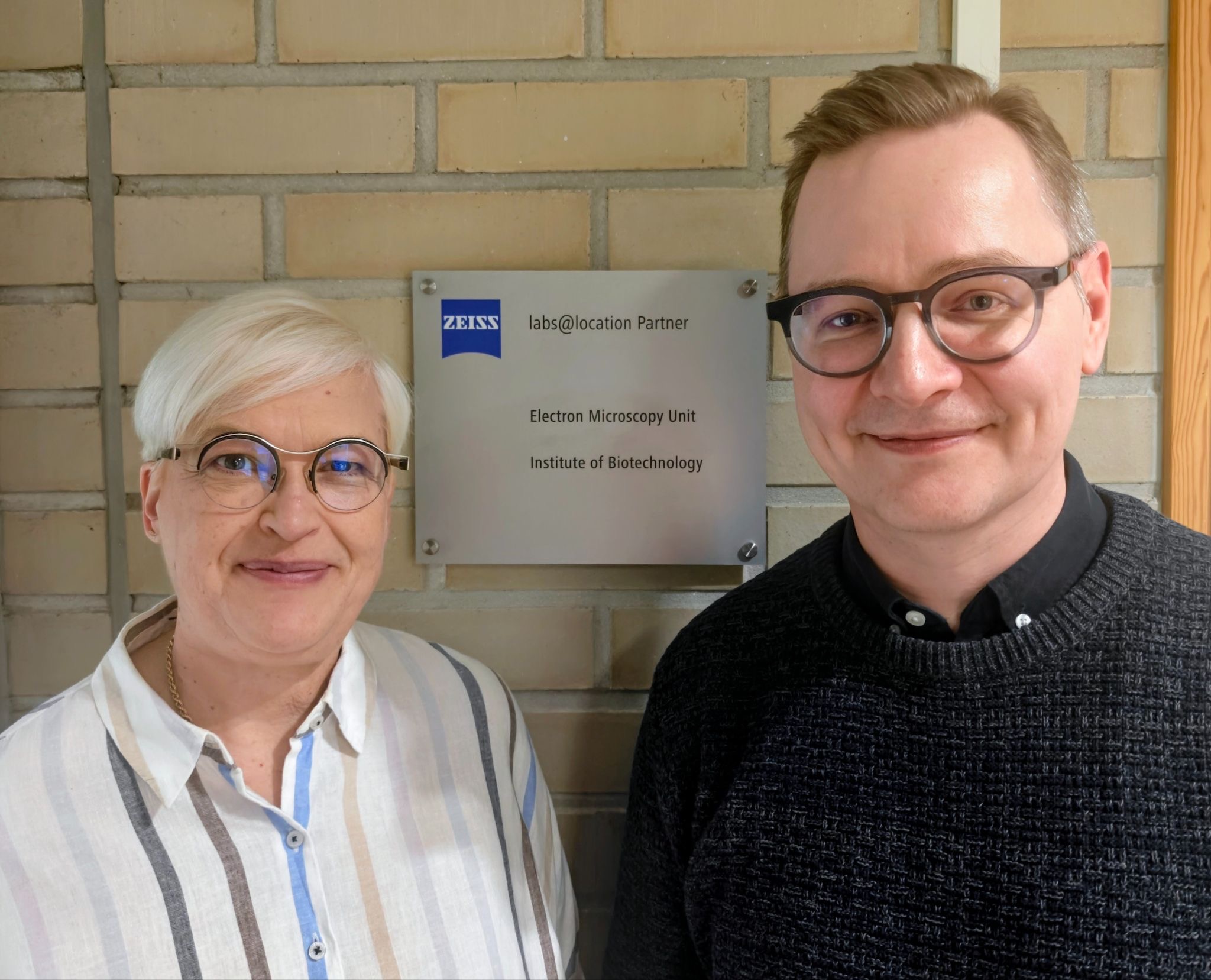
The Electron Microscopy Unit (EMBI) provides biological electron microscopy services at the University of Helsinki. Led by Research…

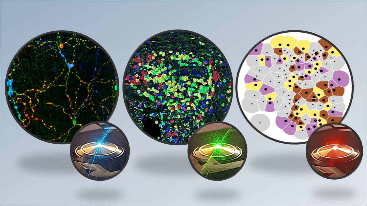
Join us for an afternoon of presentations from Euro-BioImaging users and image data/image analysis experts at our Nodes that will provide a compelling overview of the state-of-the-art in Image Data. At the Euro-BioImaging User Forum on “Image Data”, Christophe Avenel of BIIF, part of our Swedish NMI Node, shares a tool developed at the Node which provides a detailed spatial overview of MS neuropathology in both mice and humans at the single-cell level and helps shed light on the complex cellular interactions involved in the progression of MS.
What: Euro-BioImaging User Forum “Image Data”
When: March 26, 2024, from 14:00-17:00 CEST Where: Online
Abstract
Cellular architecture of evolving neuroinflammatory lesions and multiple sclerosis pathology
Christophe Avenel
BIIF, Swedish NMI Node, SE
Multiple sclerosis (MS) is a neurological disorder marked by scattered lesions and ongoing pathological changes. Insights into the disease at the cellular level have been gained through single-cell analysis, but the evolving cellular mechanisms of MS are still not fully understood. To better comprehend these dynamics, we studied the progression of neuroinflammatory lesions in mice with experimental autoimmune encephalomyelitis, focusing on changes over time and in different areas. We used In situ sequencing for single-cell spatial expression profiling, which helped us map out and categorise areas of disease activity during lesion development, revealing that active lesions expand outward. Our research shows that disease-associated (DA) glia develop separately from lesions and undergo dynamic changes throughout the disease. We also applied single-cell spatial mapping to human MS spinal cord samples, which validated the varied presence of normal and DA-glia, broke down active and inactive lesions into smaller sections, and pinpointed previously unrecognised lesion sites. Our work, which provides a detailed spatial overview of MS neuropathology in both mice and humans at the single-cell level, sheds light on the complex cellular interactions involved in the progression of MS. In this presentation, we will offer some biological context, delve into the In Situ Sequencing technology, and demonstrate how the data was disseminated for visualisation and exploration via a web portal.

April 30, 2026
The Electron Microscopy Unit (EMBI) provides biological electron microscopy services at the University of Helsinki. Led by Research…
April 29, 2026
We are delighted to announce that a new Node has joined Euro-BioImaging after stringent review by our Scientific Advisory Board and approval by the…
April 29, 2026
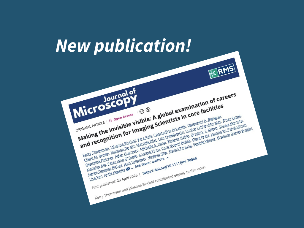
A new publication from the Global BioImaging community sheds light on the career paths, recognition, and working conditions of imaging…