
March 30, 2026
Euro-BioImaging at the EOSC ESFRI meeting in Milan
Euro-BioImaging was delighted to attend the ESFRI/EOSC Policy Workshop on “EOSC and Research Infrastructures: Opportunities and Strategies” in Milan from March 15-16, represented by…

We are delighted to announce the winners of the “Autumn” round of our Four Seasons of the Invisible Imaging Contest. Launched in early 2025, the competition highlights seasonally inspired imaging work that reveals the unseen beauty of the microscopic world. For “Autumn”, the images focus on transition and transformation, capturing a season where growth slows and structures quietly reorganise. From plants shifting from flowering to seed production to microscopic scenes echoing falling leaves and muted, earthy tones, the collection reflects autumn as a time of change, persistence, and subtle, understated beauty.
We received a high number of creative entries, and our panel of six judges had the difficult task of selecting two winners based on both artistic quality and relevance to the seasonal theme. It was a close call, with just two point between first and second place.
The first prize goes to Tomáš Figura of Czech Academy of Sciences, Institute of Botany, and Charles university for the image Is This What a Fern Looks Like?. The runner-up is Bob Asselbergh of VIB Center for Molecular Neurology, for the image Brain forests deteriorating in fall.
Tomáš will receive reimbursement of up to €1,000 to attend the scientific conference of his choice, and Bob will receive reimbursement of up to €500 to attend a scientific conference.
Check out the breathtaking winning images below and do not miss your chance to take part in the next round of Four Seasons of the Invisible imaging contest! The “Winter” round is now open for submissions until March 20.
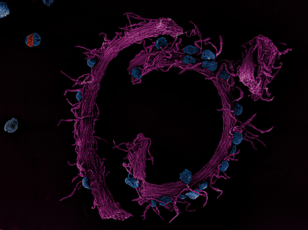
Ferns are symbols of autumn, revealing their quiet beauty when other plants have finished flowering. In regions near the equator, temperatures do not change, but humidity does. This is the best time to encounter Schizaea fluminensis, commonly known as the curly grass fern. It is a specialised species of fern belonging to the Schizaeaceae family and native to tropical regions of South America. The picture shows a purple fertile frond (sporophyll) growing at the top of the plant. The spore-containing capsules are located on the upper surface of the sporophyll and are here blue in colour. These plants are initially mycoheterotrophic and potentially mixotrophic — they steal carbon from fungi when young and possibly in adulthood too. It is a scanning electron microscopy image of gold-coated dried samples and subsequently colourised. The author acknowledges technical support from Miroslav Hyliš and Viničná Microscopy Core Facility.
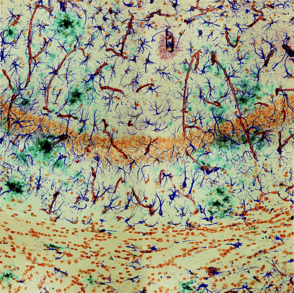
In the autumn of life, the brain enters its season of change. Neurones, once vibrant and connected, stand like thinning trees in the hippocampal forest, as Alzheimer’s plaques drift in and settle like fallen leaves. Astrocytes glow with the colours of fall, swelling and reshaping the landscape as they respond to injury and loss. Blood vessels trace winding paths through the tissue, reminiscent of streams beneath a forest floor, sustaining what remains while time slows its flow. This image captures a neural autumn — a moment where structure persists, colour deepens, and decline carries a quiet, fragile beauty. It is a fluorescence image of a 30 µm-thick mouse brain vibratome section immunostained for GFAP (astrocytes, blue), X34 (amyloid plaques, green), CD31 (dark red), and a nuclear dye (brown), acquired on a Zeiss LSM900 using a 20× Plan-Apochromat objective.
March 30, 2026
Euro-BioImaging was delighted to attend the ESFRI/EOSC Policy Workshop on “EOSC and Research Infrastructures: Opportunities and Strategies” in Milan from March 15-16, represented by…
March 30, 2026
Euro-BioImaging was delighted to attend the Public Awareness & Engagement of Research Infrastructures (PAERI) conference, represented by External Communications Officer, Marianna Childress-Poli. This year’s…

March 26, 2026
The Madrid Advanced Microscopy Center (MAdMiC) is the first Euro-BioImaging Node in Madrid (Spain). It is formed through the collaboration and close work of…