Imaging technologies support research into the structure and function of plants, shed light on plant health, resilience and adaptability, and help answer agroecology-related research questions. The Euro-BioImaging User Forum “Understanding Plant Biology” will showcase how imaging supports cutting edge research in this domain and provide information on funding opportunities for agroecology-related research projects via the AgroServ project. At this event, Clement Chambaud, CNRS, will explain how correlative light and electron microscopy in collaboration with the Bordeaux Imaging Center, part of France BioImaging, supports his research on plant tissues. Full abstract below.
What: Euro-BioImaging User Forum “Understanding Plant Biology”
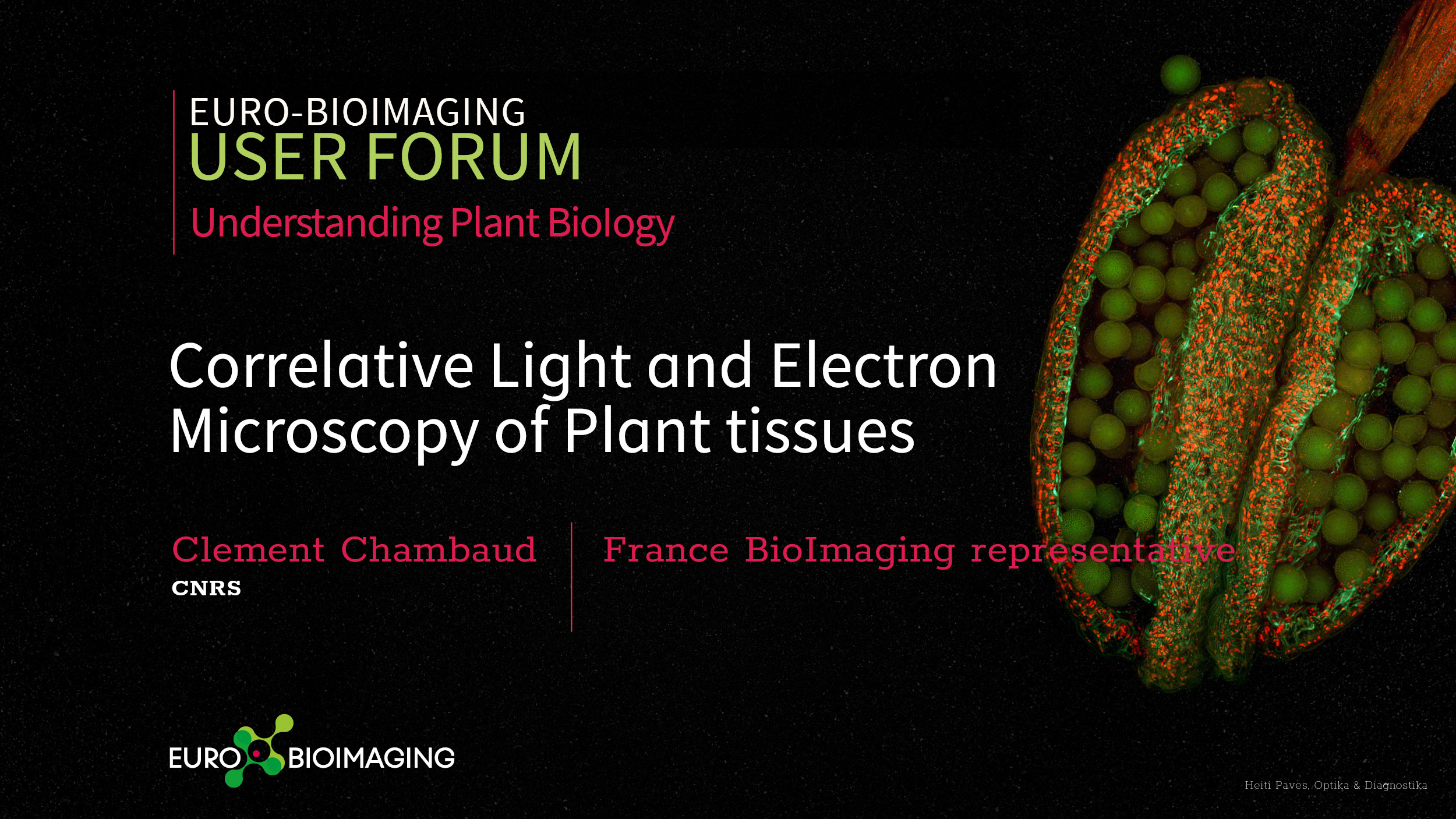
Correlative Light and Electron Microscopy of Plant tissues
Clement Chambaud, CNRS
Bordeaux Imaging Center, France BioImaging
Correlative Light and Electron Microscopy (CLEM) is a powerful imaging method that consists in combining for the very same cell or tissue area observations of light and electron microscopy. Lately, there is a growing interest in the application of CLEM in biology, especially for yeast and mammal samples, but plant applications remain scarce. Various workflows of CLEM exist and depend on the type of sample and biological questions. In this talk, Clement Chambaud will focus an approach called In Resin Fluorescence (IRF) that was adapted for plant tissues. It allows preserving the fluorescent properties of conventional fluorescent proteins during the sample preparation steps for transmission electron microscopy that, in order to have the best ultrastructural preservation possible, are usually incompatible with fluorescence preservation. CLEM protocols are always a matter of compromise between fluorescence preservation and ultrastructure. IRF for plant was adapted for roots and hypocotyl of Arabidopsis thaliana and combined with high-resolution imaging in 3D by electron tomography. It has enable us to gain insight into understanding plasmodesmata biogenesis during plant grafting, autophagy or phase separation. Here, we will develop the bottlenecks of this approach, from plant sample preparation, handling and mounting for fluorescence to transmission electron microscopy and electron tomography observations.
More news from Euro-BioImaging
April 9, 2026
Major EU funding for user access & AI development, staff training, data stewardship & many more exciting new services!
Euro-BioImaging ERIC is deeply grateful to announce that the European Union has entrusted our infrastructure with funding to shape the future of imaging and…
Euro-BioImaging is looking for an Operations Support Assistant at the Euro-BioImaging Statutory Seat in Turku, Finland, to support the day-to-day financial and administrative operations…