

March 30, 2026
Euro-BioImaging at the EOSC ESFRI meeting in Milan
Euro-BioImaging was delighted to attend the ESFRI/EOSC Policy Workshop on “EOSC and Research Infrastructures: Opportunities and Strategies” in Milan from March 15-16, represented by…
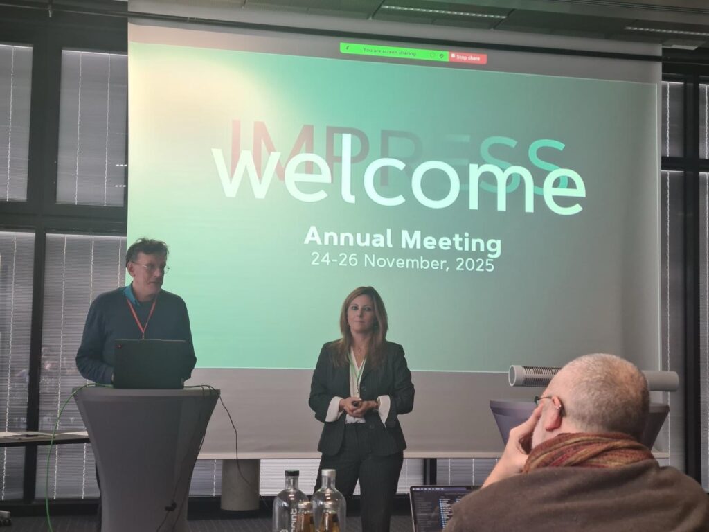
The IMPRESS project partners convened in Düren, Germany, for their Annual Meeting, bringing together scientists, engineers, and infrastructure experts for three days of technical updates and strategic discussions. Combining the SciNet Meeting with the Consortium and General Assembly sessions, the programme focused on integrating advances across the project’s Work Packages as IMPRESS moves toward building an interoperable, next-generation transmission electron microscopy (TEM) platform.
Although IMPRESS ambitious technical developments are centrally focussed on enabling novel materials science applications, its outcomes have clear potential to enrich biological EM imaging approaches. Euro-BioImaging contributes to this cross-domain exchange by bringing in the life science perspective, supporting its Node contributing to technical development for correlative imaging on biological samples, and providing training and dissemination for future users.
Bringing the life science perspective into IMPRESS
Euro-BioImaging plays a modest but meaningful role in IMPRESS: ensuring that the needs of the biological imaging community are visible as the interoperable platform and associated workflows take shape. This includes following the development of sample carriers, cryogenic approaches, and potential correlative imaging strategies that could eventually link EM innovations to biological applications.
Electron Microscopy and MassSpec Imaging experts in the team of Anjusha Mathews at Maastricht University, who are part of the AMMI Maastricht Node of Euro-BioImaging contribute to the technical development efforts, particularly where biological sample preparation and multi-modal workflows intersect with broader technical developments within the project.
One strand of the technical development work for the life science integration is focused on a cryo-microcooler that stabilises samples at low temperatures and would allow their use across different modalities, such as TEM, SEM, Raman, and AFM imaging. This demonstrates a promising step toward workflows where vitrified biological samples could be examined in high-resolution EM without losing structural integrity during transfers.
A second strand is exploring graphene monolayer liquid cells that encapsulate biological specimens in native or near-native conditions. These ultrathin graphene enclosures may make it possible to:
While still in early development, these approaches illustrate how IMPRESS technology—originally conceived for materials science—could eventually enable cross-disciplinary correlative workflows.
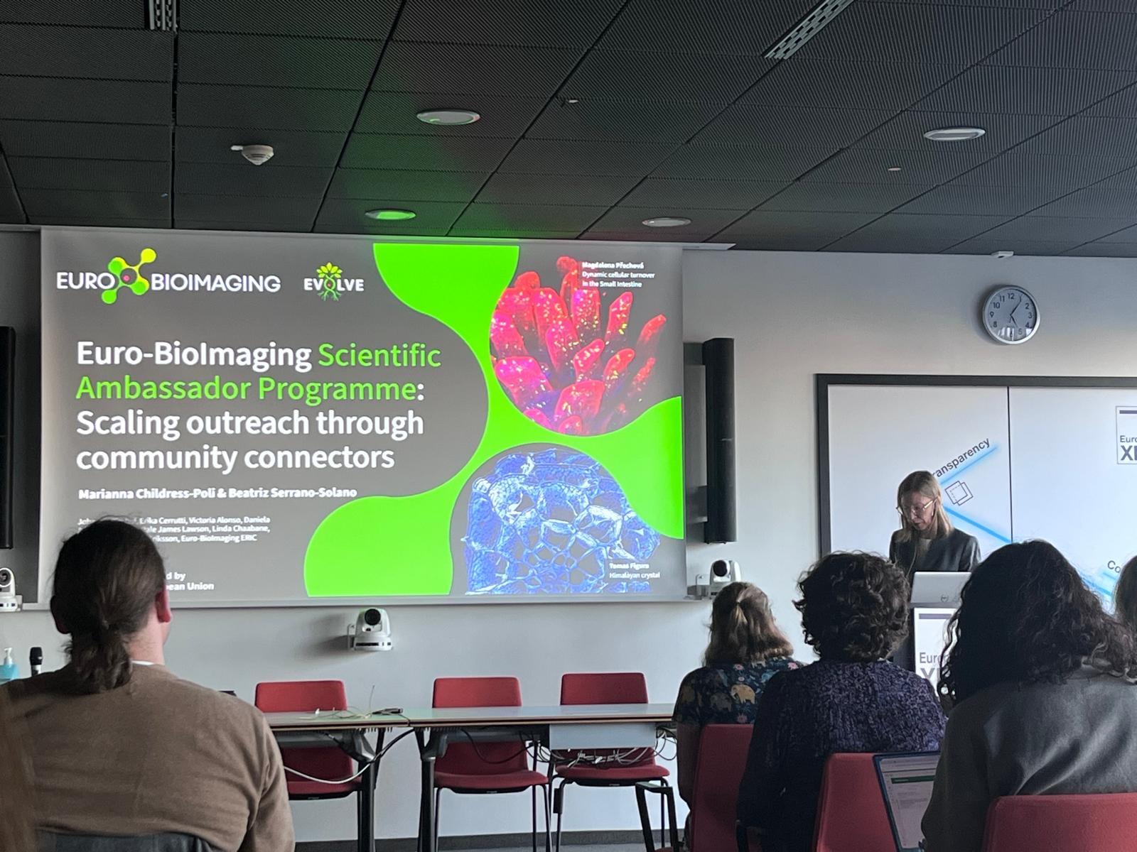
Euro-BioImaging’s contribution: training, dissemination, and community integration
Euro-BioImaging’s main involvement focuses on ensuring that results from IMPRESS can be meaningfully taken up and deployed by the wider Euro-BioImaging Node community and made accessible to life science research communities. This includes the monitoring of developments with potential relevance for biological imaging and the provision of training opportunities for RI staff on the new technologies and workflows.
The annual meeting also presented an opportunity to align with other partner Infrastructures in the proejct and well as the technical developers to plan training opportunities for 2026.
Through these activities, Euro-BioImaging ensures that the cutting-edge EM technologies developed within IMPRESS will not remain siloed in materials science and the consortium but will become valuable tools openly accessible across the research infrastructures for researchers studying biological structure, function, and dynamics.
March 30, 2026
Euro-BioImaging was delighted to attend the ESFRI/EOSC Policy Workshop on “EOSC and Research Infrastructures: Opportunities and Strategies” in Milan from March 15-16, represented by…
March 30, 2026
Euro-BioImaging was delighted to attend the Public Awareness & Engagement of Research Infrastructures (PAERI) conference, represented by External Communications Officer, Marianna Childress-Poli. This year’s…

March 26, 2026
The Madrid Advanced Microscopy Center (MAdMiC) is the first Euro-BioImaging Node in Madrid (Spain). It is formed through the collaboration and close work of…