Studying macromolecular complexes in their native environment in a cell is a real challenge, but new methods and technology advances are allowing scientists to delve into the ultrastructure of molecules even in thicker samples and study them in their context. We spoke to Simone Mattei, Team Leader for Electron Microscopy Service and Technology Development at EMBL Imaging Centre, part of our EMBL Node, where cryo-Electron Tomography, along with other novel EM approaches, are available for users via the Euro-BioImaging Proof-of-Concept study.
Tell us about Cryo-Electron Tomography imaging at EMBL IC.
Simone Mattei: My team at the EMBL IC is made up of engineers, software developers, image analysts, and molecular biologists. We are working together to develop high-throughput and fully automated pipelines to enable large-scale cryo-EM sample preparation and screening. You might be familiar with cryo-Electron Microscopy (cryo-EM). In this approach, scientists use vitrified specimens that are frozen so fast there are no ice crystals. While cryo-EM allows us to get a very high resolution view of the sample, the images we get can be approximated to projections, like an x-ray, they don’t deliver 3D information. To get a 3D model of our sample, we need to have multiple views of it. For unique objects, like cells and pleomorphic viruses, we achieve these views by tilting the stage within the microscope to image the same region of interest at different angles. We then reconstruct a 3D map of the sample, based on the thin specimens we have imaged. This is called tomography.
What are some possible applications of cryo-Electron Tomography?
Simone Mattei: The range of project applications we get for cryo-Electron Tomography/Microscopy at EMBL IC is extremely wide. Pretty much any cellular biology project could be suitable and access this technology. We see a lot of virologists now interested in how a virus replicates inside the cell. The advances in hardware and software, as well as in the sample preparation workflows, are making cryo-ET more robust and high-throughput, allowing us to apply this technique to gain insights into new fields. For large macromolecular complexes like the ribosome, we can achieve atomic level resolution in the cellular context in a wide range of samples, from parasites to marine specimens. Researchers in cellular biology should start thinking about this as a technology that is available to them and how it can push their research to a new level of resolution.
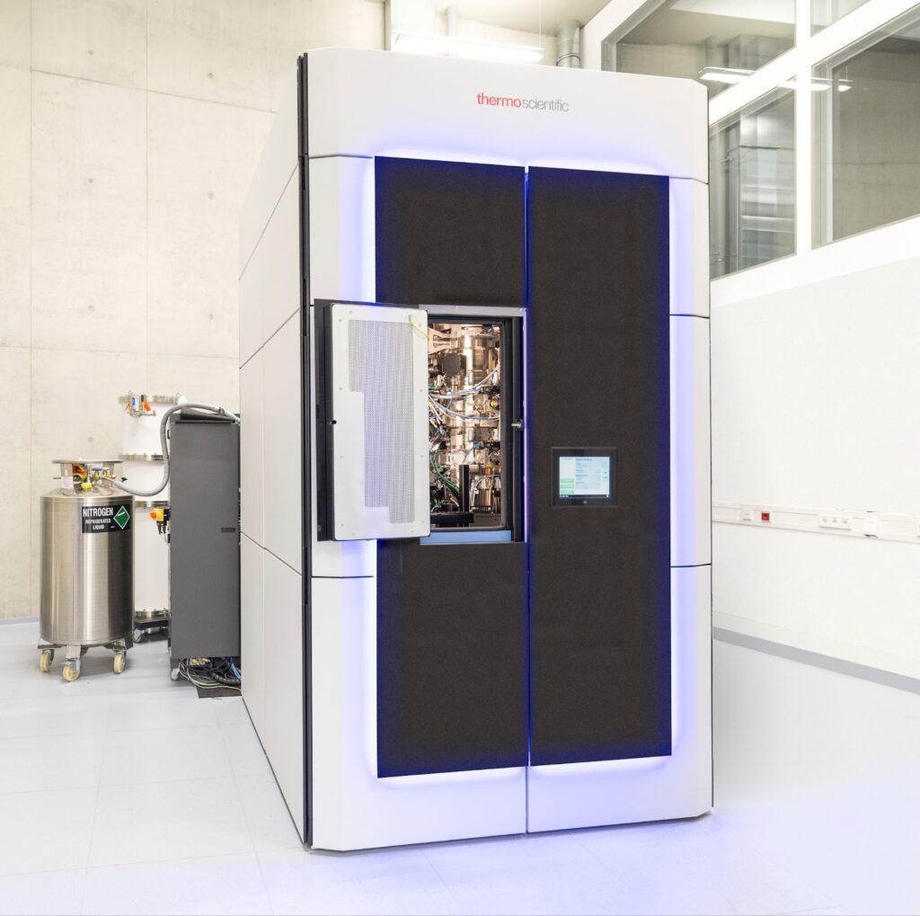
Cryo-EM instrument at EMBL IC. Photo by EMBL PhotoLab.
What kind of samples are you able to image?
Simone Mattei: Imagination is the limit. One of the major recent advancements is that we can now do tomography within thick cells, by combining Scanning Electron Microscopy (SEM) and Focussed Ion Beam milling (FIB milling). Currently we are mostly using this method on individual cells grown on special substrates, but we are working on pushing the technique further, so we can use it even on cells in their native context, so we can work with biopsies, pieces of tissues, or organoids. We can even work on whole organisms that are small enough to be vitrified by high pressure freezing.
How do you prepare an organism for an cryo-Electron tomography workflow?
Simone Mattei: We take our samples, whether organoids grown on a planchette or large specimens, into a high pressure freezer. We then get a slab of vitreous ice in which the sample is embedded. But we can’t do electron tomography with this, it’s way too thick. We have to mill away a lot of material and essentially isolate the region of interest from the thick slab of ice. The preparation step of the FIB milling is used to thin down the sample, remove cellular material from above and below the area of interest, which leaves us with a lamella is 300 nm or below in thickness.
Once we’ve created this “biopsy” in the ice, we can go with a micro-manipulator to contact the biopsy, attach the lamella to the micro-manipulator and detach it from the rest of the sample so we can lift it out - this is why the method is called "lift-out". But these lamellae are still too thick to do TEM on. So we deposit them on a receiving grid, and continue to polish them to make it thinner. This is a very challenging workflow, and our team at the EMBL IC it’s still constantly working on optimising and improving it, in order to offer it to the community.
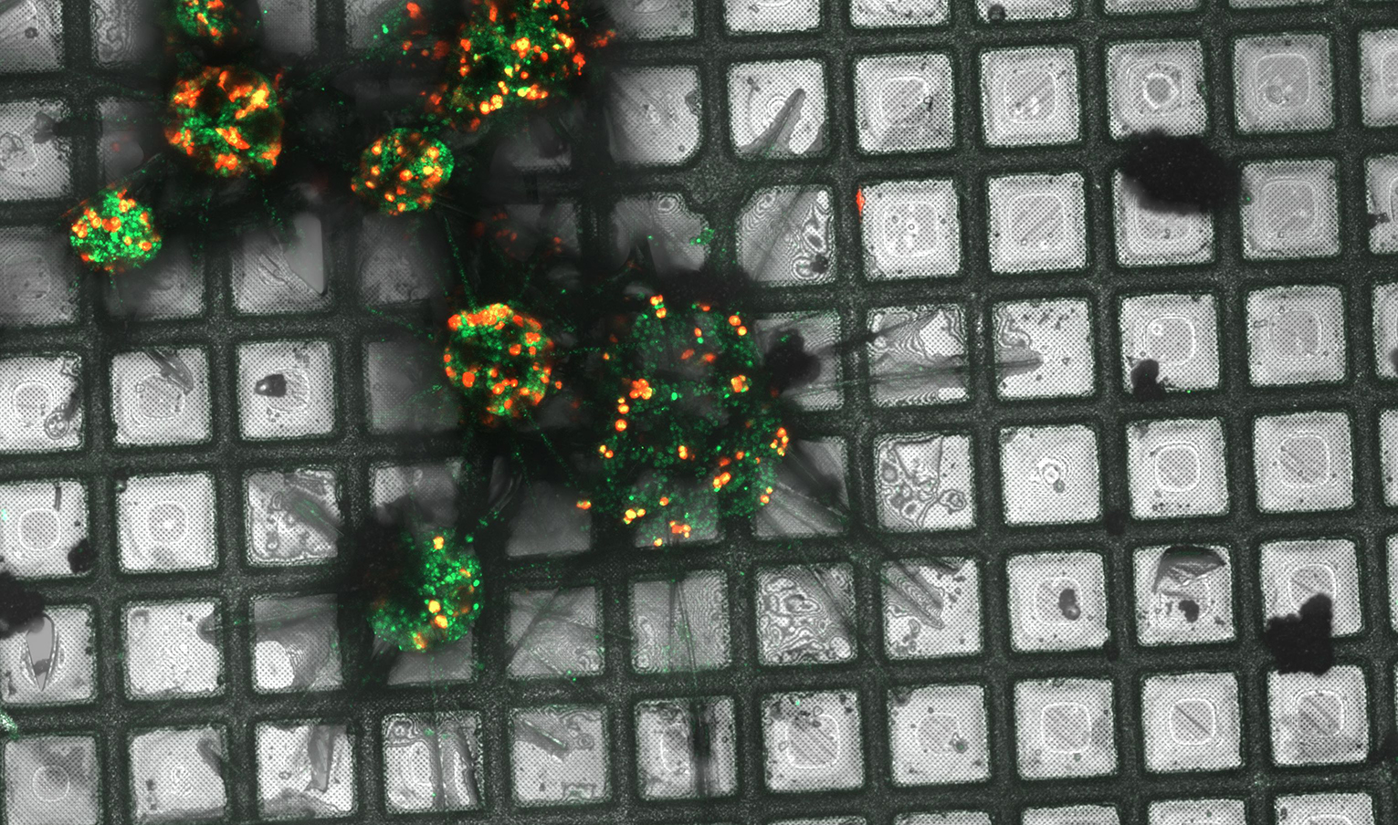
Maximum intensity projection of Acantharia (plankton), plunge frozen and imaged with a high-end confocal microscope. Green: Acantharia; red: chlorophyll of symbionts (microalgae). Credits: Anna Steyer/EMBL
What makes cryo-ET projects different from other user projects?
Simone Mattei: Service provision of these really high-tech technologies requires a lot of support from the facility. We can provide this service only under the support of our specialists. Unless a project requires a large number of visits, we generally don't train an external scientist in this technique, but we set up workshops to train the community to use the devices. We want to provide excellent support, that is our mission. We want users to get the best outcome possible.
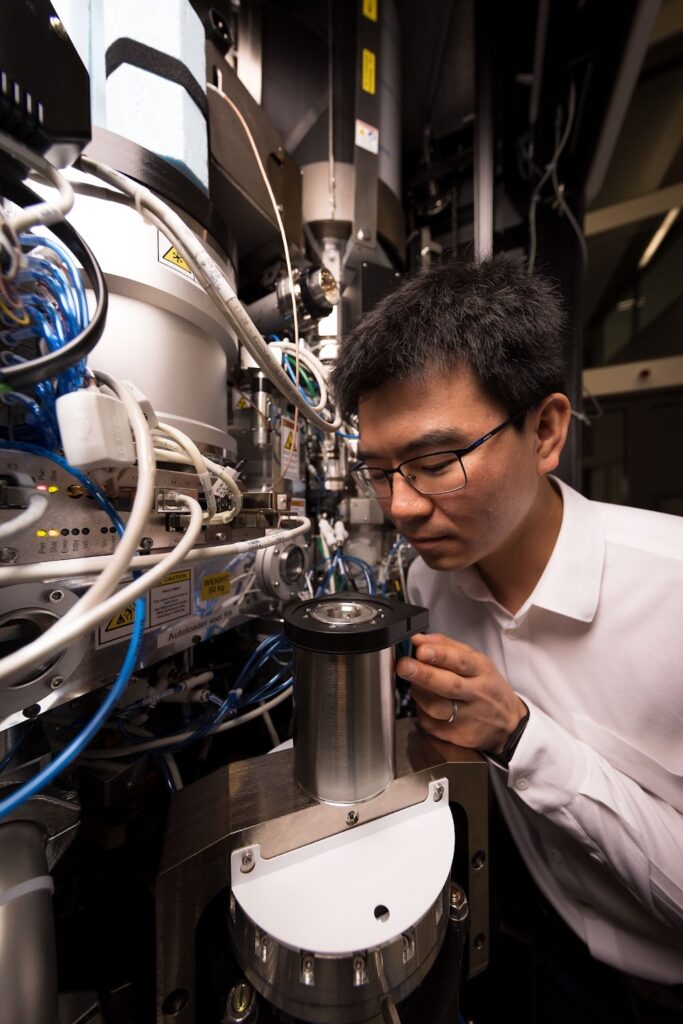
Cryo-EM expert Zhengyi Yang at EMBL IC. Photo by EMBL PhotoLab.
How does a typical project with such intense support needs usually unfold?
Simone Mattei: We have a long period of consultation before the project. We have to understand what is really scientifically sound to do, and what is technically feasible. We want the user to come with a good experimental set up and maybe even the grids with their cells or samples on them. Optimising the sample preparation and the method to locate the region of interest can take a lot of time to optimise. And then of course the actual FIB milling workflow is very time-consuming. Once we have the lamella, acquiring the tomograms does not take so much time.
Of course, once we have acquired the data, it needs to be analysed. We can support researchers with that to a certain extent. The most challenging part of the image analysis is the sub-tomogram averaging. Simply because it’s different from single particle analysis where software options are really mature. In tomography you have to jump from one software to another. Also you need a lot of computational power to perform structural studies on tomographic data
Why should users choose the EMBL IC for a cryo-ET project?
Simone Mattei: At the EMBL Imaging Centre, we give access to state-of-the-art cryo-EM equipment for structure determination projects using the latest technology and methods. For cryo-ET projects, the centre hosts two FIB-SEM devices, a Zeiss Crossbeam 550 and a Thermo Scientific Aquilos 2 equipped with an EasyLift micromanipulator and an integrated fluorescence light microscope, and two transmission electron microscopes Thermo Scientific Titan Krios G4 equipped with a cold field emission gun, a phase plate, a Selectris X energy filter and a Falcon 4i direct electron detector. Both microscopes are equipped with an automatic sample loading system and configured for automatic data acquisition. Our experts are on hand to help and support researchers during microscope handling, data acquisition and optimisation of imaging conditions.
About Euro-BioImaging’s EMBL Node
The Euro-BioImaging EMBL-Node offers a collection of state-of-the-art microscopy equipment and image processing tools. The facility was set up as a cooperation between EMBL and industry to improve communication between users and producers of high-end microscopy technology. This Node supports in-house scientists and visitors in using microscopy methods for their research and regularly organizes in-house and international courses to teach basic and advanced microscopy methods. The services provided include project planning, sample preparation, microscope selection and use, image processing and visualization. The Euro-BioImaging EMBL-Node supports advanced microscopy techniques such as FRAP, FRET, FCS, high-throughput microscopy, laser nanosurgery, CLEM, mesoscopy and super-resolution microscopy.
More news from Euro-BioImaging
July 28, 2026
Global BioImaging Advance the Conversation on Sustainable Biomedical Imaging Facilities
How can biomedical imaging facilities remain scientifically excellent while becoming more financially, environmentally, and operationally sustainable? These questions were at the heart of…
Moara Lemos: New insights from Global BioImaging’s Job Shadowing programme
Moara Lemos is a Brazilian researcher and imaging scientist. Her award winning research pushes the boundaries of structural biology, using advanced cryo-electron microscopy…
A Multiplexed Vision: Highlights from the Special Edition Virtual Pub on Spatial Transcriptomics
On June 12th, Euro-BioImaging hosted its latest Special Edition Virtual Pub, bringing together over 200 researchers and technology developers working at the cutting…