
March 26, 2026
Welcome, MAdMiC Node!
The Madrid Advanced Microscopy Center (MAdMiC) is the first Euro-BioImaging Node in Madrid (Spain). It is formed through the collaboration and close work of…

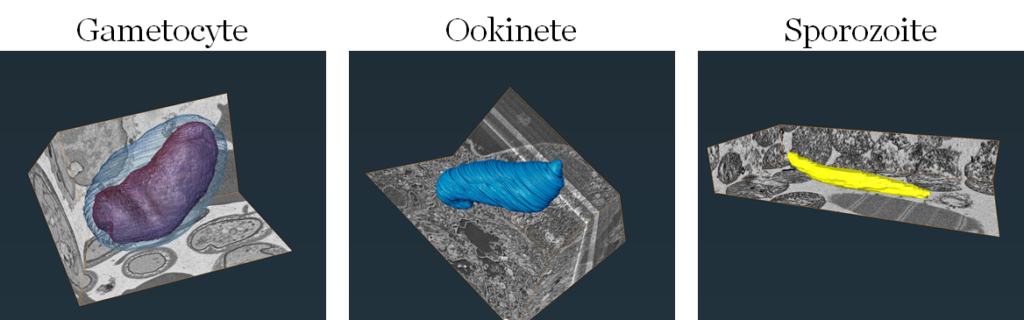
Pablo Suárez Cortés is a Postdoctoral researcher at the Max Planck Institute for Infection Biology, Berlin (Germany). His work focuses on understanding how Plasmodium falciparum, the primary human malaria pathogen, undergoes intricate transformations during its infection of Anopheles mosquitoes. In particular, Pablo aims to study secretory organelles of the Plasmodium falciparum in all three transmission stages - gametocytes, ookinetes, and sporozoites. Previously, the team identified the parasite transmission to correlate with availability of the protein PfGEST. They started exploring common functionality of this protein by means of Transmission Electron Microscopy (TEM) which, however, allowed collecting EM data from the first stage (gametocytes) only, while imaging of ookinetes and sporozoites with this technique has been challenging. Seeking expert advice and access to suitable state-of-the art EM imaging technologies, as well as funding opportunities to continue and advance his research, Pablo applied to Euro-BioImaging through the ISIDORe TNA Calls. This exciting opportunity evolved into a highly collaborative project with the Euro-BioImaging Advanced Light & Electron Microscopy Prague Node EM experts.

In his research, Pablo aims to characterize the secretory organelles during mosquito infection and investigate the role and impact of a protein of interest (PfGEST) in this process. While secretory organelles are critical components of the parasite across its different forms, the elusive nature of ookinetes and sporozoites, coupled with their inaccessibility for in vitro production, presents persistent challenges in observing and investigating these stages. Surmounting these obstacles, Pablo’s ISIDORe project strategically leverages a set of advanced EM techniques and expert support at the Electron Microscopy facility in České Budějovice, which is part of the Euro-BioImaging Advanced Light & Electron Microscopy Prague Node
Comprising Correlative Light Electron Microscopy (CLEM), 3D Serial Block-Face Scanning Electron Microscopy (SBFSEM) and Array Tomography (AT), this highly collaborative research project between Pablo and the Node EM experts team aims to gain unprecedented insights into the intricate dynamics of the secretory organelles in presence and absence (Knockout) of the protein of interest. Along this way, the challenge of studying P. falciparum ookinetes within the mosquito midgut prompted progressing developments in optimized sample preparation, imaging and analysis strategies of this biological intact system.
While the project is currently progressing and advancing, the SBF-SEM datasets have been successfully acquired for WT parasites, culminating in the generation of preliminary 3D models for all targeted stages (gametocytes, ookinetes, and sporozoites, Fig. 1). This result represents a landmark achievement in Pablo’s research and goal on P. falciparum electron microscopy imaging, particularly notable in the exploration of midgut ookinetes. Thanks to the support of the Euro-BioImaging Prague ALM & EM Node team and the ISIDORe funding - we are looking very much forward to following this project advancing and progressing towards more insights and new findings in the Malaria research field.
Article written collaboratively by Arina Rybina and Marianna Childress-Poli
March 26, 2026
The Madrid Advanced Microscopy Center (MAdMiC) is the first Euro-BioImaging Node in Madrid (Spain). It is formed through the collaboration and close work of…

March 26, 2026
German BioImaging, within its work in the NFDI4BIOIMAGE consortium, and in collaboration with Euro-BioImaging ERIC, has launched a new survey to collect input about…

March 25, 2026
Turin, Italy – 20–22 October 2026 Early career professionals working in imaging core facilities will soon have the opportunity to strengthen essential skills beyond…