On 29–30 October 2026, partners of the European PRIM-TECH3R project gathered at the University of Veterinary Medicine Vienna, Austria, for their annual project meeting. The two-day event brought together experts in disease modelling, advanced imaging and research infrastructures to discuss progress in developing complex in vitro models and preparing them for wider use by the scientific community.
Coordinated by INFRAFRONTIER ERIC, PRIM-TECH3R (Preclinical in vitro Models for the Replacement, Reduction and Refinement of Mouse Models in Preclinical Studies) aims to develop and validate advanced mouse- and human-derived in vitro models for biomedical research. By combining innovative technologies such as organoids, 3D bioprinting and organ-on-chip systems with standardised protocols and robust validation approaches, the project contributes to the 3Rs principles of replacing, reducing and refining the use of animals in research.
Euro-BioImaging is a partner in this Horizon Europe project, contributing advanced imaging technologies and expertise to support the characterisation and validation of these emerging model systems.
Developing and validating the next generation of in vitro models
A central focus of the meeting was the progress made in developing a diverse portfolio of complex in vitro models. These include lung organoids, 3D bioprinted muscle tissues, glioblastoma models, colorectal cancer organoids, skin models and blood-brain barrier systems.
While the different models are at varying stages of development, they share a common challenge: understanding how closely they reproduce the structural and functional characteristics of biological tissues and disease processes.
This is where advanced imaging plays a particularly important role. By providing detailed insights into tissue organisation, cellular interactions and physiological functions, imaging technologies help researchers characterise and validate these models, supporting their future use in preclinical research and therapeutic development.
Combining imaging expertise across Euro-BioImaging Nodes
Several Euro-BioImaging Nodes are actively contributing to the validation of models developed within PRIM-TECH3R.
At theDanish BioImaging Node, researchers are applying complementary imaging approaches, including spinning disc microscopy, multiphoton microscopy, light-sheet microscopy and high-content screening, to investigate the physiology and functional characteristics of lung organoid models.
The Advanced Light Microscopy Prague Node contributes to the characterisation of a 3D bioprinted muscle model designed to investigate cancer-associated cachexia, a condition involving severe muscle wasting. Its expertise includes optical imaging of muscle contractions and label-free nonlinear microscopy techniques, such as second- and third-harmonic generation and coherent anti-Stokes Raman scattering (CARS), providing insights into muscle structure and function.
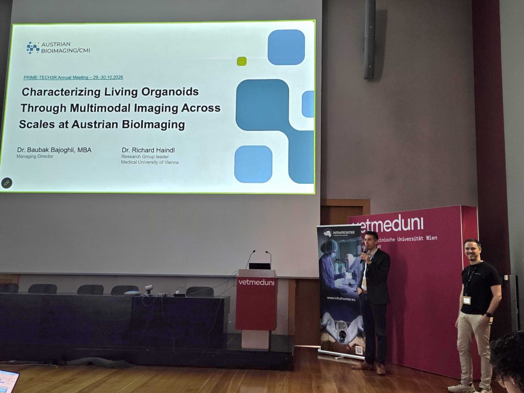
The meeting also opened new opportunities for collaboration with Austrian BioImaging, whose representatives presented a broad portfolio of multimodal and multiscale imaging technologies for organoid characterisation. Particular interest centred on optical coherence tomography (OCT), which enables non-destructive structural imaging, and its potential combination with high-resolution episcopic microscopy (HREM) for detailed three-dimensional analysis. Other approaches, including mass spectrometry imaging, offer complementary information about the chemical composition of biological samples.
Baubak Bajoghli and Richard Haindl present Austrian BioImaging's expertise at the PRIMTECH-3R event.
Discussions between the participating Nodes and model developers explored how these complementary techniques could be combined to provide more comprehensive characterisation of lung and colorectal cancer organoid models.
Connecting in vitro and in vivo imaging expertise
Beyond the characterisation of in vitro models, Euro-BioImaging also contributes expertise in in vivo imaging to support the comparison and validation of experimental models.
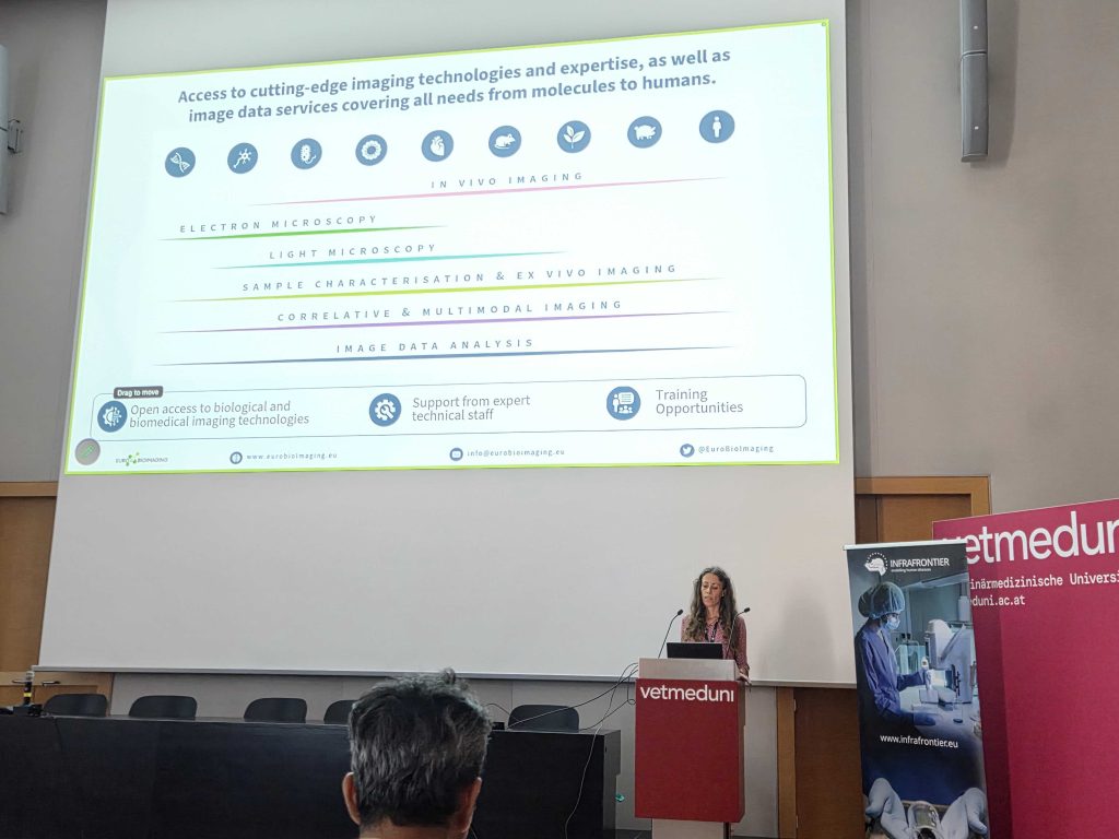
During the meeting, Erika Cerutti from the Euro-BioImaging Med-Hub presented the range of imaging services available across the infrastructure and their potential applications in the validation of preclinical models.
Erika Cerutti presents Euro-BioImaging's Technology Portfolio at the PRIMTECH-3R meeting in Vienna (November 2026). Photo courtesy of Johanna Bischof.
The discussions also highlighted some of the practical challenges of cross-border research, particularly the complex ethical and regulatory procedures associated with transferring laboratory animals between countries. These challenges underline the importance of continued European dialogue on facilitating access to specialised research infrastructure services while maintaining high standards of animal welfare.
Towards wider access to innovative preclinical models
An important objective of PRIM-TECH3R is to make its newly developed models and associated expertise accessible to the wider biomedical research community, including academic and industrial users.
The project is developing a dedicated online in vitro resource bringing together information on available models, standardised protocols and related services. Preparations are also underway for an open call that will provide potential users from industry with opportunities to access selected complex in vitro models, with the portfolio expanding as additional models become available.
The Vienna meeting demonstrated the value of bringing together complementary expertise from European research infrastructures. Through collaboration between model developers, imaging specialists and other infrastructure partners, PRIM-TECH3R is helping to establish more reliable, well-characterised and accessible experimental models for the future of biomedical research.
PRIM-TECH3R has received funding from the European Union's Horizon Europe research and innovation programme under grant agreement No. 101188158.
More news from Euro-BioImaging
November 13, 2026
Building connections for the future of Danish Life Science Research Infrastructures
How can research infrastructures build sustainable futures, strengthen collaboration and support the people who make them work? These questions were at the heart…
Following chromosomes in living cells with Oblique Plane Microscopy
How can researchers follow chromosomes over time while keeping the cells they study alive? For Rumen Stamatov, a postdoctoral researcher at Euro-BioImaging’s Sofia…