

March 26, 2026
Welcome, MAdMiC Node!
The Madrid Advanced Microscopy Center (MAdMiC) is the first Euro-BioImaging Node in Madrid (Spain). It is formed through the collaboration and close work of…
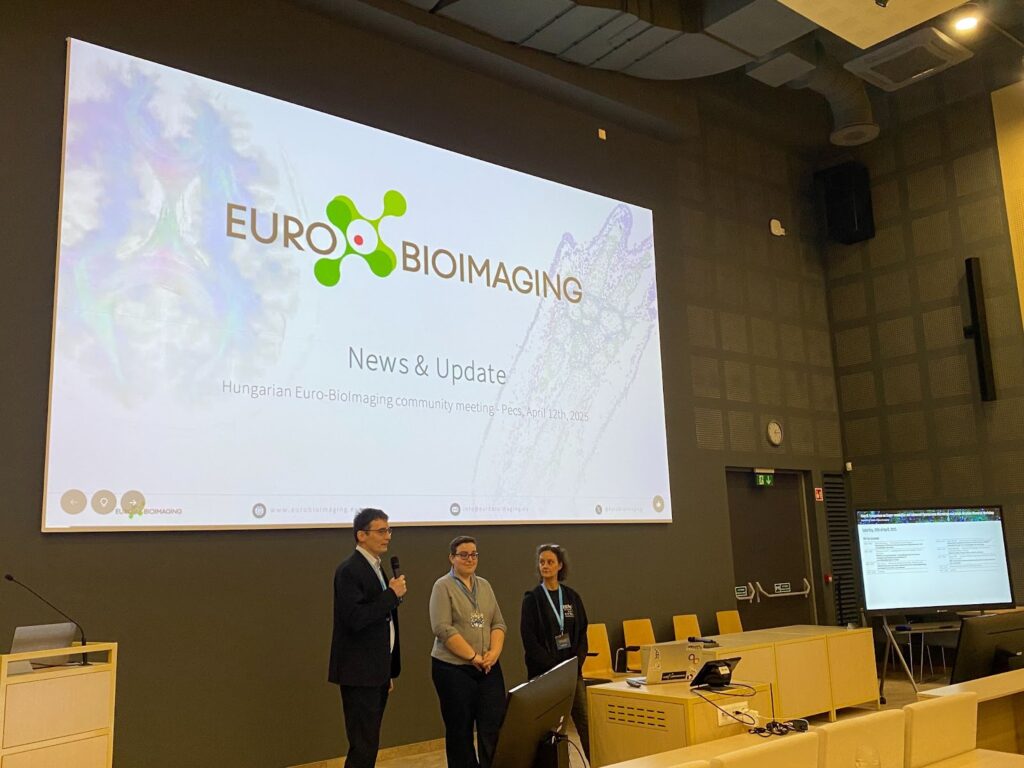
On 11-12 April 2025 Johanna Bischof (Head of Operations at Bio-Hub), Alessandra Viale (Head of Operations at Med-Hub) and Erika Cerutti (Scientific project manager) were invited to attend the István Ábrahám memorial workshop and Super-Resolution Symposium at Pécs (Hungary) with a special session bringing together the Hungarian national imaging community. This was an excellent occasion for us to meet the national imaging community, hear about new technology developments at the Hungarian Nodes, share the latest updates from Euro-BioImaging and discuss in person with the Nodes’ staff.
The Super-resolution symposium is a high level international meeting that is held annually since 2022, organised in memory of István Ábrahám, a remarkable neuroscience researcher and founder of Pécs University’s Nano-Bio-Imaging Core Facility. Former colleagues of István’s joined with national and international leading scientists in presenting their exciting research, using super-resolution technologies. The conference program featured a remarkable mix of diverse applications, focussed mainly on the areas of autism research and TRP channels, and impressed with excellent presentations.
This high-level meeting also presented an opportunity to bring together the Hungarian national imaging community. Within Euro-BioImaging, Hungary is represented with two Nodes - the Cellular Imaging Hungary Node with sites in Budapest, Debrecen, Szeged, and Pécs; and the Hungarian Medical Imaging Node with sites in Budapest, Debrecen, and Pécs.
On Saturday 12th, the special imaging community session featured talks from representatives from many of the Node sites and from Euro-BioImaging, providing an important platform for sharing updates and to facilitate exchange.
Alessandra and Johanna presented the opportunities that Euro-BioImaging can offer for both researchers and Euro-BioImaging facility staff. Particular attention was dedicated to the EVOLVE training, Job shadowing and mentoring program opportunities and to the funding opportunities for user access, such as the canSERV open call. With positive experiences as hosts of an ISIDORe-funded user access project, and an already awarded canSERV project, canSERV was seen by the audience as a great occasion to benefit from the funds available at Euro-BioImaging to apply for access to imaging technologies.
The Hungarian Nodes were well represented by several staff members, several who travelled across Hungary to Pecs only for the afternoon session, and together with the researchers who attended the conference they were actively engaged in discussion and learning more on the Euro-BioImaging opportunities.
The different Node sites presented their latest technology developments, workflows, and innovations. The impressive diversity of topics and tools included, among others:
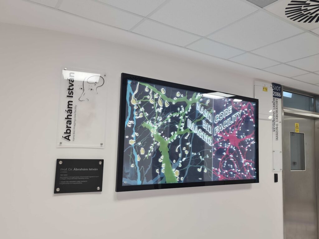
Being onsite at the University of Pécs also presented the opportunity to visit the Nano-Bio-Imaging Core Facility and meet the local facility staff. The facility features an impressive range of advanced microscopes, focussed on super-resolution systems, including STED, 3D single-molecule detection, STORM, and SIM.Beyond super-resolution, the facility also has an Stimulated Raman Spectroscopy system and a platform for laser dissection and capture, and provides support for its users with image analysis, experimental planning, and sample preparation.
The custom-built and designed rooms of the facility provide optimal conditions to perform high-end imaging experiments and reveal the experience and expertise of István Ábrahám, who championed and designed the facility, although he unfortunately never got to see it in full function.
The Hungarian Medical and Preclinical Imaging Node is a multi-sited, multimodal Node covering biomedical imaging both at preclinical and medical level. The Node sites are based in Debrecen, Pécs and Budapest and being invited at the István Ábrahám memorial workshop was a good opportunity to also visit some of the Node’s facilities.
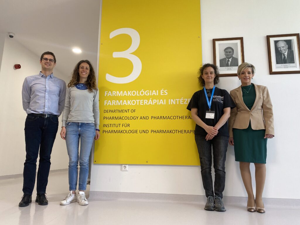
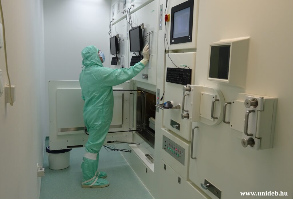
At the Debrecen facility, Erika and Alessandra met Arató Viktória, node manager, where she presented the main research pipelines ongoing at the Department. They are equipped with a 16,5 MeV cyclotron GMP certified for the production of PET radiopharmaceuticals: [18F]FDG, [18F]FET, [11C]MET and [68Ga]PSMA, which are also distributed to external Institutes across Hungary and Romania, and with a micro-PET that can be used for metabolism and biodistribution studies. They are involved in several research topics mainly focussed on angiogenesis studies, creation of animal models and the development of cyclodextrin based agents for radiotherapeutics delivery.
Once in Pécs, they met Zsuzsanna Helyes and Ádám István Horváth, part of the staff working at the Preclinical Imaging Platform at Pécs University. It was established in 2012 at the department of Pharmacology and Pharmacotherapy of the Medical School and it is extremely well equipped with cutting-edge preclinical imaging instruments including a luminescent and fluorescent in vivo optical imaging system, suitable for functional imaging, a fluorescent molecular tomograph, a laser speckle imaging system for the real time measurement of blood perfusion changes in tissues and an in vivo micro-CT for the visualisation of structural alteration of the bone microarchitecture, lung structural changes, emphysema and fibrosis and body fat composition. This equipment provides valuable tools for investigating pathophysiological processes and identifying novel drug targets in inflammatory diseases.
Beside these advanced in vivo imaging equipment, they also have several laboratories and spaces in support to the in vivo imaging activities such as cell culture and animal facilities equipped with a huge number of tools for preparing and testing diverse models of inflammation and neuropathies.
This article was written collaboratively by Erika Cerutti and Johanna Bischof.
March 26, 2026
The Madrid Advanced Microscopy Center (MAdMiC) is the first Euro-BioImaging Node in Madrid (Spain). It is formed through the collaboration and close work of…
March 26, 2026
German BioImaging, within its work in the NFDI4BIOIMAGE consortium, and in collaboration with Euro-BioImaging ERIC, has launched a new survey to collect input about…
March 25, 2026
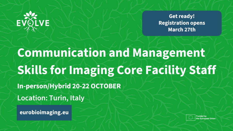
Turin, Italy – 20–22 October 2026 Early career professionals working in imaging core facilities will soon have the opportunity to strengthen essential skills beyond…