How can researchers follow chromosomes over time while keeping the cells they study alive? For Rumen Stamatov, a postdoctoral researcher at Euro-BioImaging’s Sofia BioImaging Node in Bulgaria, an Imaging 4 All grant made it possible to set up a protocol to explore a new imaging approach at Euro-BioImaging’s EMBL Node, gaining valuable experience along the way.
Rumen studies how chromosomes move and behave throughout the cell cycle, combining microscopy with machine learning and physical modelling. At his home institute, he works extensively with Airyscan microscopy, using deep learning to reduce noise in images acquired with low laser power. This helps address a central challenge in live-cell imaging: collecting enough detail without exposing cells to damaging levels of light.
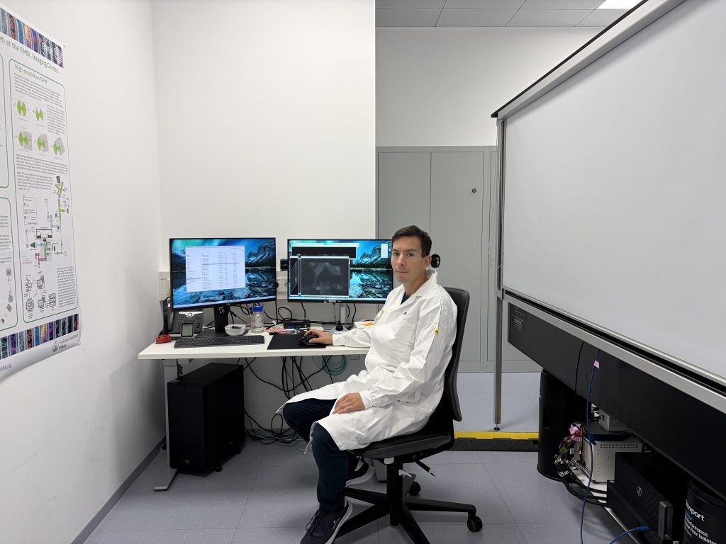
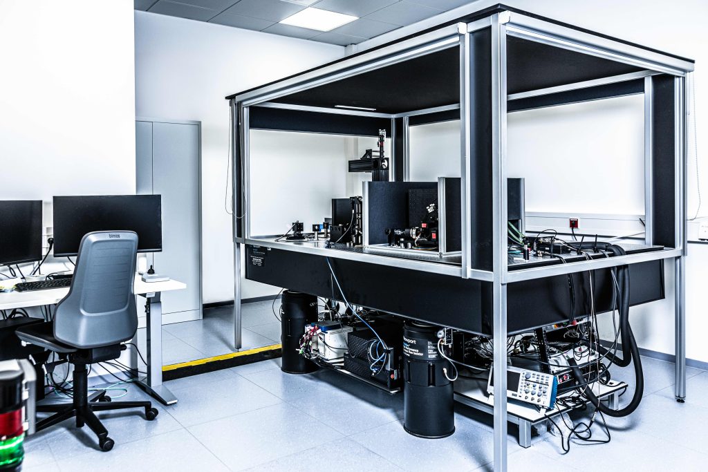
Through the Imaging 4 All programme, Rumen visited EMBL to work with a custom-built oblique plane microscope, a system unavailable at his home facility. Its light-sheet illumination offered an opportunity to image living cells more gently and capture their structures in three dimensions.
Finding the right balance
Transferring an experiment to a new microscope can be more difficult than it his sounds. After his cells arrived at EMBL, Rumen spent two weeks working with the team to optimise the imaging conditions, followed by a week of experiments.
His patience paid off. In one experiment, he acquired images every five minutes over 48 hours while keeping the observed cell alive—an encouraging demonstration of what carefully optimised imaging conditions could achieve.
“As a microscopist, it is important to have experience in different systems. And you have to learn patience. That is the weapon to get through,” says Rumen.
Bringing experience back to Sofia
The visit gave Rumen practical experience in adapting his samples and experiments to a different imaging system. He is interested in exploring whether a similar microscope could eventually be built in Bulgaria with technical support, making the knowledge gained at EMBL relevant to future developments at his home facility.
For Rumen, the exchange was also a reminder of the importance of continued learning, even for experienced microscopists. In the accompanying video, he shares his experience and invites researchers interested in live-cell imaging, chromosomes and single-cell projects to collaborate with the Sofia BioImaging Node.
Imaging 4 All is coordinated by Global BioImaging and funded by Wellcome, supporting access to imaging technologies, training and expertise.
Rumen Stamatov at the EMBL Imaging Facility where he performed live cell imaging with Oblique Plane Microscopy.The Oblique Plane Microscope at the EMBL Imaging Centre. Photo by EMBL/PhotoLab, used under a CC-BY-IGO license.
More news from Euro-BioImaging
November 9, 2026
First NL-BioImaging Advanced Image Analysis Course brings Dutch bioimage analysts together
From 21 to 24 September, Erasmus MC in Rotterdam hosted the first NL-BioImaging Advanced Image Analysis Course. Supported by NL-BioImaging and the NVvM,…
EVOLVE Course: Communications and Management Skills for Imaging Core Facilities
20–22 October 2026 | Turin, Italy | Hybrid course The Communications and Management Skills for Imaging Core Facilities course brought together 50 participants…