June 3, 2026
BMIC meets Flanders BioImaging
In May 2026, the Belgian Molecular Imaging Community (BMIC) and Flanders BioImaging joined forces in Ghent for a two-day community event that brought…

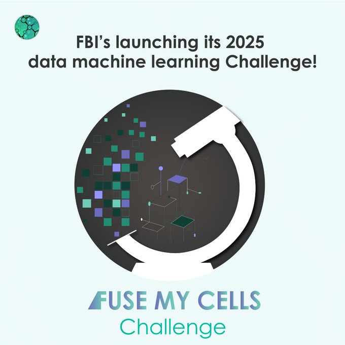
Hey, deep learning experts! Show off your 3D image-to-image fusion skills using deep learning by entering France-BioImaging‘s Fuse My Cells challenge.
The main objective of the Fuse My Cells challenge is to predict a fused 3D image using only one or two available 3D views, providing a practical solution to the limitations of current microscopy techniques, such as improving image quality, extending the duration of live imaging, saving on the photon budget, and facilitating image analysis.
How to participate?
The competition is divided in two phases:
You have until the end of the first phase, on February 28, 2025, to register and participate at this Fuse My Cells challenge. Nonetheless, you can start working on your preliminary algorithm and tests on January 31st, 2025 (with the release of the training database)!
For top 3 winners:
You can visit the original article (from which this text was borrowed) on the France-BioImaging website.
June 3, 2026
In May 2026, the Belgian Molecular Imaging Community (BMIC) and Flanders BioImaging joined forces in Ghent for a two-day community event that brought…

June 1, 2026
The EOSC Association has adopted a new position paper calling for a distinct Work Programme-based Partnership for EOSC in FP10. Euro-BioImaging welcomes this…
May 29, 2026
Spatial transcriptomics is rapidly transforming the way researchers study complex biological systems. To explore the latest developments in this fast-moving field, Euro-BioImaging is…