
March 30, 2026
Euro-BioImaging at the EOSC ESFRI meeting in Milan
Euro-BioImaging was delighted to attend the ESFRI/EOSC Policy Workshop on “EOSC and Research Infrastructures: Opportunities and Strategies” in Milan from March 15-16, represented by…

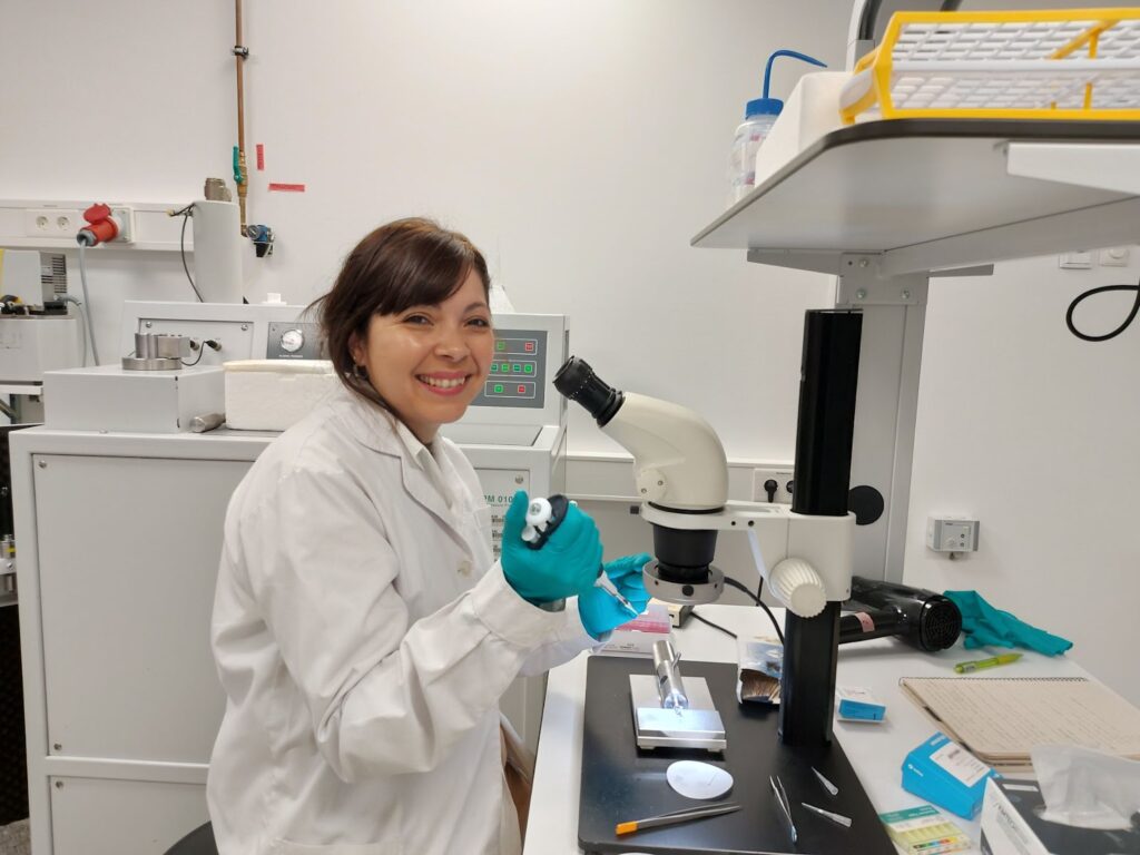
The Euro-BioImaging EMBL Node is happy to welcome Fátima Silvina Galván, a researcher at the Laboratorio de Microbiología Ultraestructural y Molecular del Centro Integral de Microscopía Electrónica (CIME), a research unit of Consejo Nacional de Investigaciones Científicas y Técnicas (CONICET) and Universidad Nacional de Tucumán (UNT), in Tucumán, Argentina. Her visit is funded by Global BioImaging’s Imaging 4 All initiative, which supports researchers from low- and middle-income countries (LMICs) in accessing advanced imaging technologies.
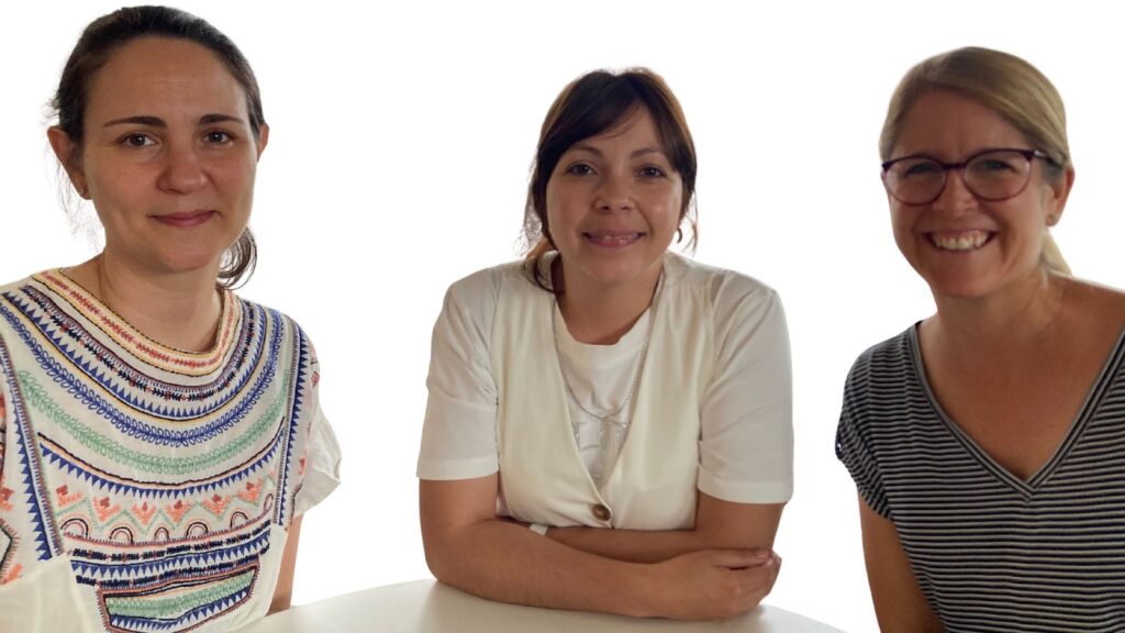
Silvina, together with the scientific team led by Dr. Virginia Albarracín, an Independent Researcher at CONICET and Director of CIME-CONICET-UNT, dedicates her work to studying microbial populations adapted to extreme ecosystems. Their research focuses on communities thriving in lakes and salt flats of the Argentine Puna (HAAL), a high-altitude region of the Andean plateau (between 3,000 and 6,000 meters above sea level).
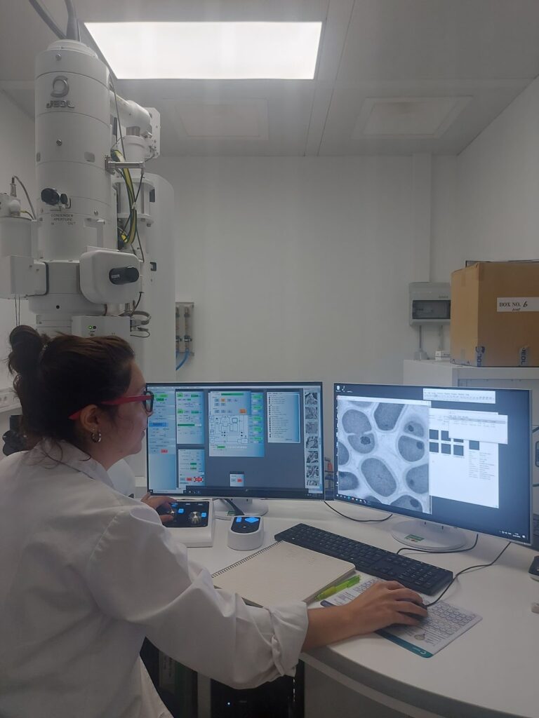
Specifically, Silvina concentrates on understanding and elucidating, through genomic approaches and ultrastructural studies using advanced microscopy techniques, such as scanning electron microscopy (SEM) and transmission electron microscopy (TEM), the adaptation mechanisms of Exiguobacterium sp. S17. This polyextremophilic bacterium was isolated from stromatolites, which are stratified microbial communities that form macroscopic columnar structures at the sediment–water interface and are characterized by a high mineral content.
Exiguobacterium sp. S17 is notable for its ability to survive —and even thrive— under extremely harsh conditions, including toxic concentrations of arsenic, high UV radiation, elevated salinity, and reduced atmospheric partial pressure of O₂. These traits make it a strong candidate for bioremediation purposes, for instance, to remove soil pollutants, with potentially significant positive impacts on society.
Previous studies on S17 have revealed its capacity to produce nanotubular structures that serve as physical communication systems between neighboring bacteria, enabling survival and adaptation in response to high UV radiation doses. This discovery, reported for the first time in the Exiguobacterium genus and in extremophilic bacteria from the HAAL, is documented in the following article: https://pubmed.ncbi.nlm.nih.gov/39964557/
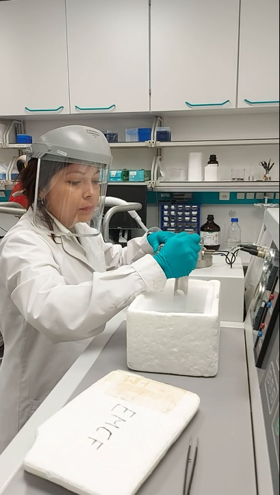
At EMBL’s Electron Microscopy Core Facility, Silvina will learn new Electron Microscopy approaches, such as high-pressure freezing, that will allow her to study the Exiguobacterium without chemical fixation, and explore the nanotubular structures in high resolution.
Fascinated by this extraordinary form of bacteria and curious to help Silvina find an answer to her scientific question, the team of EM experts led by Rachel Mellwig, Operations Manager at EMBL’s EMCF, have helped Silvina to master the high pressure freezing approach and guided her so she can perform 3D electron tomography to study where the tube is connecting to the cell. At EMBL, Silvina will also perform fluorescence microscopy experiments with Beate Neumann and the Advanced Light Microscopy Facility (ALMF) team to see if there is the transfer of material between the nanotubes connecting the bacteria. If this is confirmed, correlated light and electron microscopy pipelines could be established to target specific events.
I would like to sincerely thank the programme funded by Global BioImaging’s Imaging 4 All initiative for allowing me to turn this project into reality and for placing trust in both my academic background and the feasibility of the proposal. This stay has been a truly enriching experience, both academically and culturally.
-- Silvina Galván
During her two month stay at EMBL, Silvina will learn multiple new imaging methods, many of which she can apply in her lab in Argentina.
“The project is really inspiring,” says Rachel. “Together, we will gather as many observations as possible, which Silvina and her colleagues can analyse at home.”
“I am deeply grateful to Dr. Yannick Schwab and Rachel Mellwig, who, from the very beginning, when we proposed this project together with Dr. Virginia Albarracín, were fully supportive and willing to help us with both the planning and the application process for the Global BioImaging fellowship. I would also like to sincerely thank the programme funded by Global BioImaging’s Imaging 4 All initiative for allowing me to turn this project into reality and for placing trust in both my academic background and the feasibility of the proposal. This stay has been a truly enriching experience, both academically and culturally. I consider it highly successful, as I was able to accomplish the goals I had set for this period. I’m delighted to have worked at EMBL Heidelberg,” says Silvina, with a smile.
We are really excited to see how this project progresses, and pleased to see how open-access imaging infrastructures can foster global scientific exchange.
March 30, 2026
Euro-BioImaging was delighted to attend the ESFRI/EOSC Policy Workshop on “EOSC and Research Infrastructures: Opportunities and Strategies” in Milan from March 15-16, represented by…
March 30, 2026
Euro-BioImaging was delighted to attend the Public Awareness & Engagement of Research Infrastructures (PAERI) conference, represented by External Communications Officer, Marianna Childress-Poli. This year’s…

March 26, 2026
The Madrid Advanced Microscopy Center (MAdMiC) is the first Euro-BioImaging Node in Madrid (Spain). It is formed through the collaboration and close work of…