Uriel Koziol, a professor at the Universidad de la Republica, Montevideo, Uruguay, studies parasitic tapeworms (such as Echinococcus and Taenia), a type of worm that causes serious disease in humans and livestock. Despite the threat these organisms pose to human & animal welfare, very little is known about the first larval stage of the life cycle, when it can infect a human host. Indeed, the larval organism is so small that it is impossible to study its detailed anatomy with a traditional confocal microscope. To gain insight into this critical period, Uriel Koziol applied to use Serial Blockface Scanning Electron Microscopy at the Euro-BioImaging’s Finnish Advanced Microscopy (FIAM) Node in Helsinki. In this story, Uriel Koziol explains how this state-of-the-art imaging technology has allowed him to better understand the life cycle & larval anatomy, without ever leaving his home country.
“Studying the life cycle of Echinococcus is very difficult. We want to better understand the larval phase – when it is infectious to humans. How does it enter the intestine? How does it carry out infection?” says Uriel Koziol. “But, this is nearly impossible because it is too dangerous. So we work with a model organism called Hymenolepis microstoma. It is very similar to Echinococcus and Taenia on the cellular and basic biology level, but poses no risk to human health.”
With this model, Uriel and his team are starting to better understand the anatomy of the larva. They have identified 30 cells, including muscle cells and nerve cells, and have noticed the nuclei in these cells disappearing. “Why are the nuclei disappearing? The larvae are so small, it’s very difficult with confocal microscopy to resolve this problem,” says Uriel.
Need for high-end Electron Microscopy
Uriel knew that Electron Microscopy (EM) would bring better resolution. But he suspected that figuring out the distribution of the cells, and how they are organized in the larva would be practically impossible with normal EM methods, because it would take too much time. But then, Uriel read about a special EM method to do serial sections in the literature.
“For the resolution we want, Serial block-face scanning electron microscopy is really the only option.”
Potential funding opportunity
At around the same time, Uriel learned about the Euro-BioImaging pilot User Access fund from an email circulated by Latin America BioImaging, platform for the bioimaging community across Latin America that is part of the Global BioImaging network, like Euro-BioImaging. He looked at the Euro-BioImaging website and saw that the Finnish Advanced Microscopy Node in Helsinki offered the special type of Electron Microscopy he was looking for and in addition, had data processing experience. He sent in his application and was overjoyed when his project was awarded funding – with one condition. He would have to carry out the project remotely – in order to stay within the allotted budget.
“Once the project was accepted, we started meeting with the team in Finland to see how we could implement the project, from sample preparation to data analysis. The node sent me protocols about how to fix the samples, which we did here in Uruguay. To be on the safe side, we prepared more samples than would be needed,” says Uriel. While shipping samples is very expensive and complicated, it remained the best solution to fit the budget. “And once the samples are prepared,” continues Uriel, “they can be preserved for a long time.”
Working remotely
During the pandemic, the Finnish ALM Node implemented a number of processes and technical solutions to enable virtual microscopy (learn more in this article). Uriel’s project was therefore not the first remote project the Node had undertaken, and Uriel was very happy with the whole process. “From the beginning, there was a lot of communication with the colleagues in Finland and I always felt secure,” explained Uriel. “For example, when the Node received the package they called me and sent a picture, signaling a sample that may have come into contact with air. Together, we chose which samples to put on the microscope, what regions to look at, and also made decisions about the angle of view. Despite the time difference, I was able to be present at every imaging session.”
“And Ilya Belevich from the Node has been extremely helpful with data processing. This was really important for me. You really have to know about the technology in order to interpret the data. There can be artefacts, you have to be aware of it.”
Serial block-face scanning electron microscopy is a very powerful technique and perfectly adapted to Uriel’s scientific question. To date, he has completed 7 imaging sessions, in total 5-10 imaging sessions will be needed to complete his mission. With time, he is confident he will be able to reconstruct the whole larva. This is an amazing outcome, and something he could have only dreamed of, without open access to imaging infrastructures provided via Euro-BioImaging.
Developing larva of Hymenolepis microstoma. Image by Uriel Koziol, Ilya Belevich, and Eija Jokitalo.
More news from Euro-BioImaging
June 12, 2026
Euro-BioImaging joins Canadian scientific platform community for CNSP 10th anniversary meeting in Ottawa
Euro-BioImaging, represented by Antje Keppler, Director or the Euro-BioImaging Bio-Hub, was delighted to join the Canadian Network…
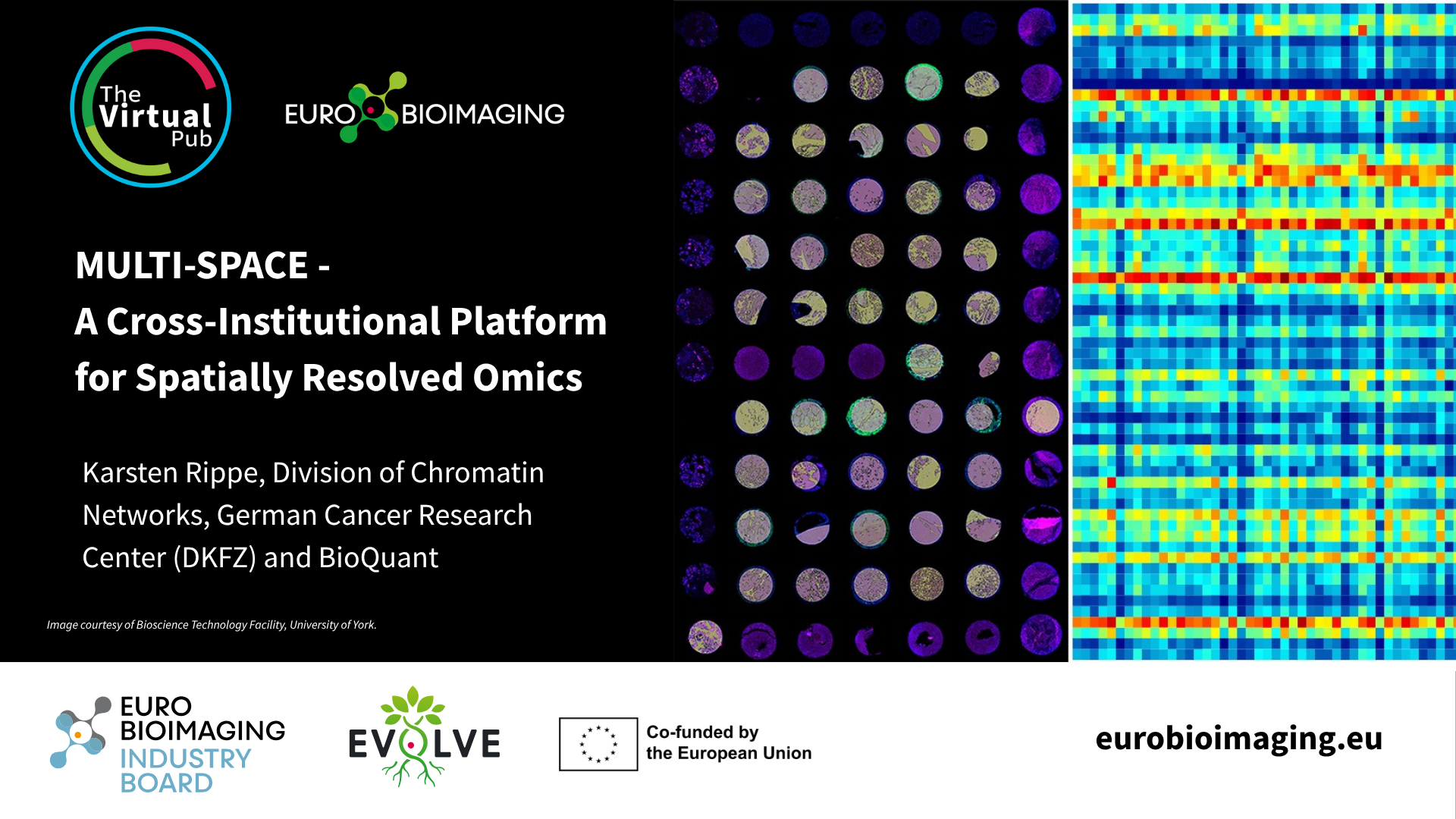
MULTI-SPACE - A Cross-Institutional Platform for Spatially Resolved Omics
Spatial transcriptomics is rapidly transforming the way researchers study complex biological systems. To explore the latest developments in this fast-moving field, Euro-BioImaging is…
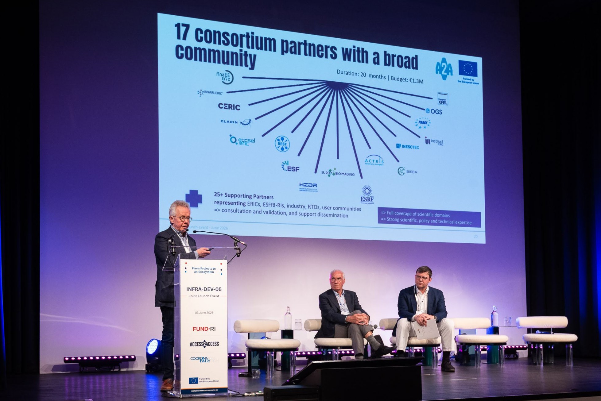
From Projects to an Ecosystem: Launching a Shared Vision for the Future of Europe’s Research and Technology Infrastructures
On 3 June, colleagues from across Europe’s research and innovation community gathered in Brussels to launch three Horizon Europe initiatives that will help…