In this study, all assay development, sample preparation and microscopy were performed at Heidelberg University’s Infectious Diseases Imaging Platform (IDIP) in biosafety containment 2 and 3 conditions (BSL‑2 and 3). The EMBL-Node contributed to the development of an automated image analysis workflow as part of the semi-quantitative high-throughput microscopy-based assay. Its mission - in collaboration with the Kreshuk lab at EMBL - was image and data handling, analysis and visualisation. Specifically, the goal was to identify human serum that contained SARS-CoV-2 specific antibodies (one of the three major classes present in human serum, IgG, IgA and IgM), and to develop a computational approach to carry out the identification process for a high volume of samples. In other words, the basic approach was to use cells infected with SARS-CoV-2 as an analysis platform to test whether serum prepared from human blood samples contained antibodies reacting with the viral antigens present on the infected cells.
Data management
The amount of data handled in the initial two procedures was large – up to 50.000 images on 50 plates. Data processing time was between 2-3 hours per plate. Over the course of the study, up to 4 TB of data were processed and analyzed. The ALMF’s extensive experience in the analysis of high-throughput microscopy screens, the image analysis expertise of the Kreshuk Lab, as well as EMBL’s high-performance computational resources were very important to this study.
The assay was then deployed in a large-scale (5,000 volunteers) population immunity study in Baden- Württemberg, Germany, conducted by a consortium of regional hospitals. The objective was to test for virus immunity in pairs of children and parents before reopening local schools. High-throughput microscopy and semi-automatic image analysis were used to identify specific SARS-CoV-2 antibodies in the study patients. To find out more about the methods and results of this important study, read the preliminary study report.
An effective procedure for diagnostics
The study showed that the sensitivity and specificity of COVID-19 diagnosis - as compared to an approved ELISA-based diagnostic test - is improved with high-throughput microscopy and an automated image analysis workflow. One of the advantages of the method is the presence of all viral antigens on the SARS-CoV2-infected cells, as compared to only one (or a selected few) in ELISA-based and similar diagnostic tests. As different persons may develop different spectra of antibodies against the virus, the presence of all viral antigens (instead of one or a few), reduces the chances for false negative readings. It provides a compelling argument for using microscopy for COVID-19 diagnosis – and for the serological analysis of other virus infections.
Sharing COVID-19 data sets with other researchers
To ensure the data developed in this study benefits a maximum number of researchers, all raw microscopy images, intermediate segmentation and infected cell classification results from this study, as well as quality control and final score results are available in the BioImage Archive (http://www.ebi.ac.uk/bioimage-archive) under accession number S-BIAD24. We hope you will have a look!
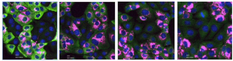
Figure 1: Virus-infected cells employed in assays to test for the presence of SARS-CoV2-specific antibodies in human blood samples. Example images with immunolabeled SARS-CoV2-infected human cells showing the blood serum response of four different persons to the virus infection (Pink: virus RNA; Green: IgG antibody; Blue: Cell Nuclei). The leftmost image shows a very strong correlation of IgG binding with virus infection, indicating a strong immune response. The two central images show a weaker response. The right-hand image shows no reactivity. The rightmost image is from blood serum that was acquired before 2018 (pre-Covid19) and consequently shows no response.
About Euro-BioImaging’s EMBL Node
The Euro-BioImaging EMBL-Node offers a collection of state-of-the-art microscopy equipment and image processing tools. This Node supports in-house scientists and visitors in using microscopy methods for their research and regularly organizes in-house and international courses to teach basic and advanced microscopy methods. The services provided include project planning, sample preparation, microscope selection and use, image processing and visualization. Through Euro-BioImaging, life scientists, regardless of their affiliation or area of expertise, can apply to access services from 21 internationally-renowned imaging facilities called Nodes.
About the BioImage Archive The BioImage Archive stores and distributes biological images that are useful to life-science researchers. Its development will provide data archiving services to the broader bioimaging database community. This includes added-value bioimaging data resources such as EMPIAR, Cell-IDR and Tissue-IDR.
More news from Euro-BioImaging
July 28, 2026
Global BioImaging Advance the Conversation on Sustainable Biomedical Imaging Facilities
How can biomedical imaging facilities remain scientifically excellent while becoming more financially, environmentally, and operationally sustainable? These questions were at the heart of…
Moara Lemos: New insights from Global BioImaging’s Job Shadowing programme
Moara Lemos is a Brazilian researcher and imaging scientist. Her award winning research pushes the boundaries of structural biology, using advanced cryo-electron microscopy…
A Multiplexed Vision: Highlights from the Special Edition Virtual Pub on Spatial Transcriptomics
On June 12th, Euro-BioImaging hosted its latest Special Edition Virtual Pub, bringing together over 200 researchers and technology developers working at the cutting…