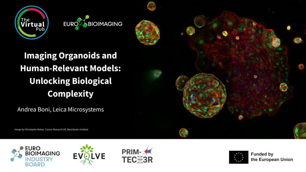
We will host the Special Edition Virtual Pub "Imaging Organoids" on Friday, January 30, from 1-3 pm CET. It will focus on methods used to image organoid systems. As part of this event, Andrea Boni, Leica Microsystems, will give a talk entitled "Imaging Organoids and Human-Relevant Models: Unlocking Biological Complexity" (description below). Join us to hear this and other exciting presentations introducing a range of methods used to image organoid systems - from lung buds to mini-brains.
Title: Imaging Organoids and Human-Relevant Models: Unlocking Biological Complexity
Presenter: Andrea Boni, Leica Microsystems
Organoids and human-relevant models are transforming biomedical research by providing physiologically accurate systems that closely resemble human tissue architecture and function. Microscopy-based imaging is essential for unlocking the full potential of these models, enabling high-resolution visualization of cellular organization, dynamic processes, and disease phenotypes in a 3D context.
In our presentation, we will showcase application examples where the Viventis Deep, our open-top dual-view and dual-illumination light-sheet microscope, was used to study the development and morphogenesis of intestinal organoids. We will highlight how advanced imaging approaches allow researchers to capture intricate structural details and dynamic cellular interactions over time. In addition, we will discuss some of Leica’s latest innovations in organoid imaging, including solutions designed to improve throughput, image depth, and compatibility with complex 3D models, ultimately accelerating discoveries in developmental biology and disease modelling.
More news from Euro-BioImaging
April 7, 2026
canSERV User Meeting highlights impact and future perspectives for cancer research in Europe
The canSERV Annual Meeting, held in Brussels from 25–27 March 2026, brought together researchers, service providers, Research Infrastructures, policymakers, and patient representatives to reflect…
We were delighted to take part in the 21st European Molecular Imaging Meeting (EMIM), held in Ljubljana, Slovenia, from March 24–27, 2026. The conference…
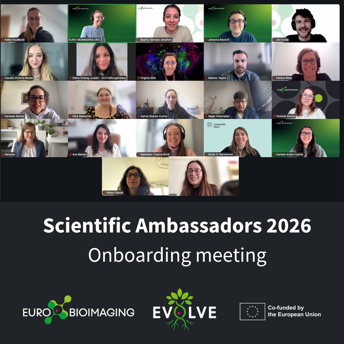
Euro-BioImaging Welcomes its 2026 Scientific Ambassadors Cohort
Following the continued success of the Scientific Ambassadors programme, Euro-BioImaging is delighted to welcome its 2026 cohort. Building on the strong foundation laid by…