Would learning to use the most cutting-edge imaging instruments help you answer your scientific question? Could world-class technology accelerate your research? We spoke toNádia Nascimento da Rosa, a PhD student at the Pequeno Príncipe Faculties and Pelé Pequeno Príncipe Research Institute in the State of Paraná, Brazil, whose neurodegenerative disease research project was boosted when she became a Euro-BioImaging user at the Facility of Multimodal Imaging - AMMI Maastricht Node. With funding from the Brazilian government, Nádia embarked on a six months scientific journey that would shape not only her PhD project but her whole career. We spoke to Nadia to find out more about how this Euro-BioImaging user project opened the door to high-tech international collaboration and impacted her scientific results.
Nádia Nascimento da Rosa is a PhD student in Brazil. She works as a bioprocess engineer and biotechnologist, in the Advanced Therapy and Cellular Biotechnology in Regenerative Medicine Research, in a group led by the neurologist Professor Katherine Athayde Teixeira de Carvalho. The group’s focus is on the use of Mesenchymal stem cells for neurodegenerative diseases.
Bioprinting to support a novel therapy option
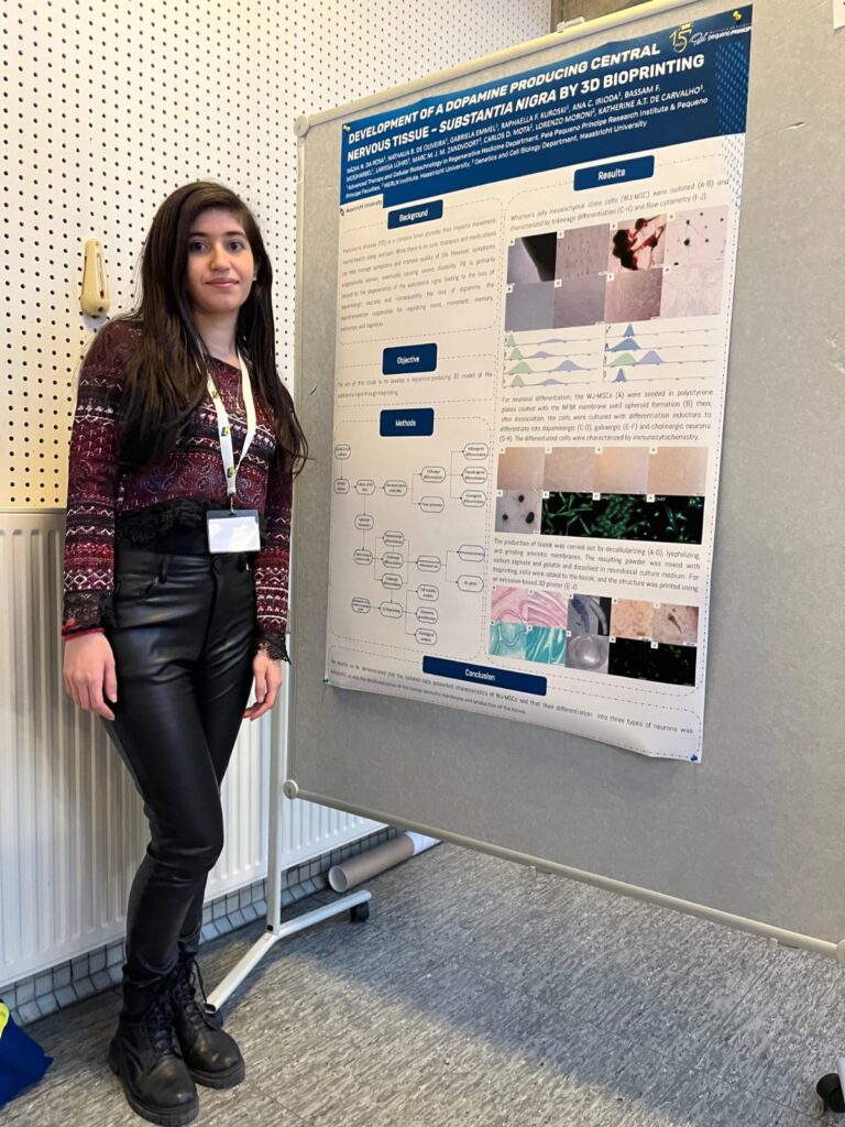
“My project is the development of a 3D structure, through bioprinting, similar to the substantia nigra, which is the part of the brain affected in Parkinson’s Disease. And for that I’m differentiating Mesenchymal stem cells into neural precursor cells (immature neurons), then dopaminergic, cholinergic and gabaergic neurons, which are the types of neurons present in the substantia nigra. Then, using a hydrogel based on amniotic membrane and an extrusion based bioprinter, I print these neurons into a 3D structure. The goal is that this structure will be functional and will produce dopamine,” explains Nádia.
“But I ran into a wall at my home institute, because we only have one fluorescent microscope. With this microscope, it is impossible to study a thick structure like the one I developed,” she explains.
Embarking on an international collaboration
Supported by her Professor, who has a long-standing collaboration with Marc van Zandvoort at Euro-BioImaging’s AMMI Maastricht Node, Nádia applied to use the confocal and two-photon microscopes at the AMMI Maastricht Node as a Euro-BioImaging user. These state-of-the-art microscopy approaches would allow her to see her whole structure in 3D and also look at live cells. To fund her visit, Nádia turned to the “CAPES” programme, a scholarship from the Brazilian government that enables Brazilian doctoral students to do a doctoral internship abroad. The CAPES funding would give her the funds necessary to stay six months in the Netherlands, a true scientific journey with an important impact on her research and her career.
When Nádia arrived in Maastricht, she was amazed by the state-of-the-art imaging equipment that was available in the lab. She had absolutely no experience with two photon microscopy but Marc and his team provided hands on individual training she needed to work independently with her samples. In addition, she participated in the M4I – Microscopy Autumn School – an imaging course run by the Node, where she was exposed to many different microscopy approaches.
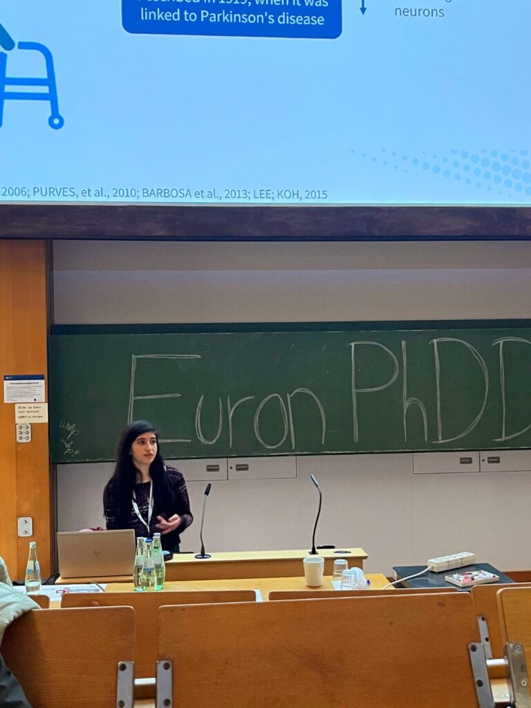
Nádia's scientific journey in Europe included attending the EURON PhD Days in Cologne, where she had a poster presentation and an oral presentation.
Taking advantage of state-of-the-art technologies
Before leaving Brazil, Nádia prepared her samples, so she could work with the same cells she was working with in her lab. “I cultivated the mesenchymal stem cells and prepared the amniotic membrane, then my colleague sent them to me in the Netherlands by mail. But to complete my experiment, I needed access to a bioprinter in Maastricht.”
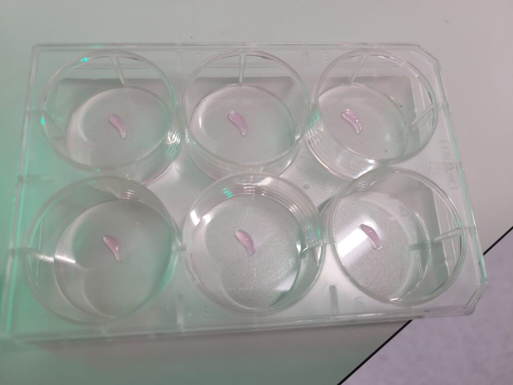
Bioprinted structures that Nadia works with. Image courtesy of Nádia Nascimento da Rosa.
So, Marc arranged a collaboration with Professor Lorenzo Moroni and Professor Carlos Mota, of the MERLN Institute, a centre for regenerative medicine at the Maastricht University. “The bioprinting in Maastricht was quite different from what I have in my home institute,” explains Nádia. “Working with these cutting-edge machines was a real game-changer for me.”
Exciting results
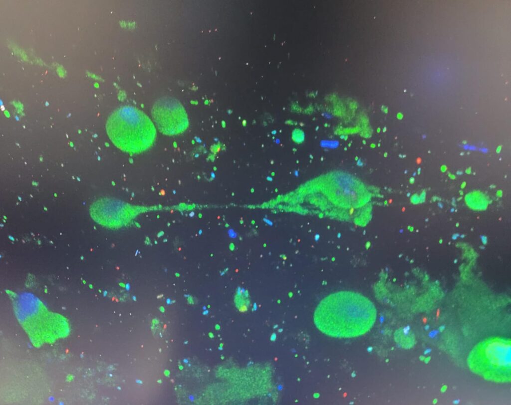
With her new structures and with the power of two-photon and confocal microscopy, Nadia proceeded with her experiments. Today, she is back in Brazil, and she is extremely happy with all the data she has to analyse. In particular, she’s happy with the images she obtained – to show the viability of the cells, the cytochemistry (analysed with the confocal microscope), as well images of the cells inside the hydrogel, attached to the membrane and connected to it.
Looking at the neuronal cells in the hydrogel with two-photon microscopy. Image courtesy of Nádia Nascimento da Rosa.
“The images are beautiful - and they prove that the cells are still expressing the neuronal protein,” says Nádia. “Now I must test to see if the genetic expression of the cells is still the same, and analyse to see if the cells are producing dopamine.”
“Going to Europe was eye-opening,” says Nádia. “I learned so much. In addition to the rich collaboration with colleagues in Maastricht, I got to give an oral presentation at the EURON PhD Days, a congress for neurosciences that took place in Cologne. I also really enjoyed visiting a photonic microscopy lab at Aachen University. But the most amazing outcome of this experience is that my professors have started the process that will allow me to graduate with a double PhD, from Pelé Pequeno Príncipe Research Instituteand the University of Maastricht,” says Nádia with pride.
This six-month scientific journey has been a life-changing experience for Nádia. It has allowed her to build lasting collaborations with Professors Lorenzo Moroni, Carlos Mota and their team at MERLN around 3D printing and with Professor Marc van Zandvoort and his team for imaging. In the long-term, Nádia hopes to translate this research into an actual treatment for Parkinson’s disease. In the meantime, her story is a compelling example of how excellent science brings people together all around the globe. Euro-BioImaging is proud to provide for collaboration, ensuring that all researchers have access to the state-of-the-art imaging technologies they need to answer their scientific questions.
More news from Euro-BioImaging
April 9, 2026
Major EU funding for user access & AI development, staff training, data stewardship & many more exciting new services!
Euro-BioImaging ERIC is deeply grateful to announce that the European Union has entrusted our infrastructure with funding to shape the future of imaging and…
Euro-BioImaging is looking for an Operations Support Assistant at the Euro-BioImaging Statutory Seat in Turku, Finland, to support the day-to-day financial and administrative operations…