
Lattice Light-sheet: For fast, subcellular, volumetric imaging
February 28, 2023
LLSM
Proof-of-Concept
Swedish NMI Node
Want to see adherent cells with low phototoxicity and photobleaching? What about spheroids and organoids? Both of these are possible with Lattice Light-sheet imaging, an interesting approach for those studying adherent cells, membrane systems, spheroids and organoids. We spoke to Steven Edwards, at the Advanced Light Microscopy facility at SciLifeLab in Sweden, part of our Swedish NMI Node. His facility - as well as others within Euro-BioImaging - are accepting applications for Lattice Light-sheet projects as part of our Proof-of-Concept study. Read more about this technology and what it can be used for in the interview below.
We are today talking about Lattice Light-sheet imaging. Please provide a short summary of this type of imaging and list some applications:
My name is Steven Edwards, and I provide scientists with light-sheet and lattice light-sheet support at the Advanced Light Microscopy facility at SciLifeLab in Stockholm, Sweden. We were fortunate to receive a beta version of the Zeiss Lattice Light-sheet 7 in summer 2020. The microscope is ideal for fast, subcellular, volumetric imaging of adherent cells with low phototoxicity and photobleaching. It is also possible to image 3D cell cultures such as spheroids and organoids as well as cells growing within a hydrogel matrix.
Standard sample carriers, such as coverslip-bottom petri dishes and multi-well chambered coverslips are ideal for parallel imaging experiments with multiple treatment conditions. For the best performance, care should be taken to use high-precision No. 1.5 coverslips.
Soon, our beta microscope will be replaced with a serial version which has an additional sCMOS camera for simultaneous two-color imaging, improving the time-resolution further. The new cameras will have improved low-light performance which will allow us to further reduce the light dose to the sample.
Due to the volumetric imaging speed, the microscope is also suitable for high-throughput imaging of model membrane systems.
Tell us a bit more about a specific project that was done in your facility using this technology? What scientific questions were you addressing?
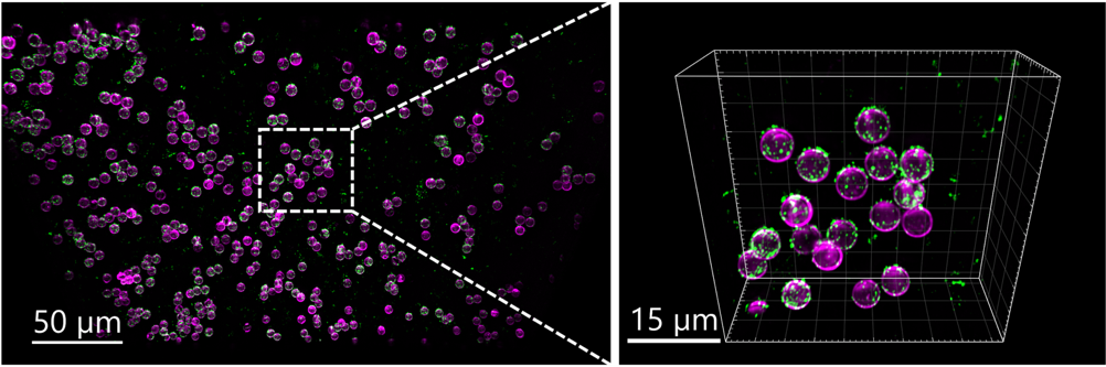
The plasma membrane of cells is highly complex, consisting of thousands of lipids and proteins, complicating the study of host-virus interactions. Three-dimensional model membrane systems allow us to create simplified models of the plasma membrane with precise control of the lipids and proteins it is composed of. In a recent preprint, published by Jan Schlegel et al., 5 μm diameter silica beads were coated with a lipid bilayer and angiotensin converting enzyme 2, to screen for unknown host-virus interaction partners. Read the scientific article here.
What are some advantages of this technique that make it suited to addressing this type of question?
Lattice Light-sheet microscopy made it possible to perform fast, volumetric imaging of virus-like particles binding to proteins on the surface of membrane coated beads. Image stacks were acquired of thousands of beads, orders of magnitude faster than a conventional point-scanning confocal microscope. Lattice Light- sheet microscopy allowed us to image with minimal photobleaching, which is particularly problematic for small, delicate samples such as virus-like particles. In addition, motion artifacts which were visible in point-scanning confocal stacks were reduced.
What other services do you provide in your facility that would be useful in combination with this type of imaging?
Users that want to perform live cell imaging with higher resolution are instead directed towards our Zeiss Elyra 7 platform which provides fast super-resolution imaging. For larger model organisms, such as zebrafish, the Zeiss light sheet Z.1 is the instrument of choice. The two Light- sheet systems complement one another and cover a resolution range suitable for imaging subcellular organelle dynamics up to centimeter sized mouse brain hemispheres.
We offer full support for all user projects including methodological setup (e.g. design of study protocols and standard operating procedures), technical assistance to run instruments as well as quantitative image analysis of the often huge 4D data sets generated by these techniques.
How to apply:
Lattice Lightsheet is part of the Euro-BioImaging Proof-of-Concept study. The Proof-of-Concept study makes it possible to introduce exciting, new imaging technologies to our portfolio that were previously unavailable via our network. We are currently accepting applications to use these technologies at participating Nodes as part of the Proof-of-Concept study. Be part of this study - and contribute to community-wide continuous technological innovation!
All scientists, regardless of their affiliation, area of expertise or field of activity can benefit from Euro-BioImaging’s pan-European open access services. Potential users of these new technologies are encouraged to submit project proposals via our website. To do so, you can log in to access our application platform, choose the technology you want to use and the facility you wish to visit, then submit your proposal. All applications will be processed by the Euro-BioImaging Hub. As usual, users will benefit from advice and guidance by technical experts working at the Nodes, training opportunities, and data management services.
For more information: info@eurobioimaging.eu
More news from Euro-BioImaging
