Katerina Kaduchova is a PhD student at the Centre of Plant Structural & Functional Genomics, Institute of Experimental Biology, Czech Academy of Sciences at Olomouc. She studies the fundamental processes that regulate plant growth in crop plants, particularly in barley. In October 2023, she attended a two-day course on “Multi-modal light microscopy imaging in plant research,” organized by Katerina Malinska, IFIEB in Prague, part of Euro-Bioimaging’s Advanced Light & Electron Microscopy Prague Node. During this workshop, she tried out, several microscopy systems, experimented with root/growth tracking software, and discovered how Virtual Reality can contribute to image analysis.
Every year, Katerina Malinska with the IFIEB team organizes a course on Light microscopy imaging in plant research, intended for plant researchers, PhD students and highly motivated pre-graduate students. This workshop allows participants to compare the light microscopy modalities available at IFIEB, in particular for live-cell confocal fluorescence microscopy.
Exploring cereal crops’ genomes
“We want to explore the organization and dynamics of cereal crops’ genomes. Until now, we have primarily focused on easy-to-image parts of the barley plants such as the roots. In my home institute, we use confocal microscopy with horizontal sample placement. But we were very curious about the non-invasive, long term, live-cell imaging optimized for plants our colleagues in Prague do with vertically mounted samples. We’ve been in contact with them. The training course was the perfect opportunity to meet them in person and get hands on experience,” says Katerina Kaduchova. (Learn more about Katerina’s work in her recent publication: https://onlinelibrary.wiley.com/doi/10.1111/tpj.16355 )
“Prior to the course, we prepared our fluorescent marker lines, for tracking of the dynamics of the nuclei and cells in barley,” says Katerina.
Trying new microscopes and software
At the course, she used a confocal microscope with a vertical sample mounting, and a special root tracking software, that gave her new insight into her sample and made the time-lapse analysis more efficient.
“It was great to get hands on training with this really nice software for tracking roots. In addition, the experts showed me new possibilities on how to mount my samples, which was extremely helpful,” she explains.
She also tried the ultrafast simultaneous two-channel imaging on spinning disk microscope, SRRF, perfusion chamber. “This was really amazing,” says Katerina. “Especially in combination with the two fast cameras. For us, it is particularly interesting to see super-fast cellular processes in combination with the tracking.”
An intensive course that includes some fun
Katerina Kaduchova was one of 19 attendees at the workshop. “We strive for an informal, friendly atmosphere,” says the course organizer, Katerina Malinska. “The two-day workshop has four parallel hands on sessions. Each session has only 5 participants , so it’s very intense. But we also try to include some fun.”
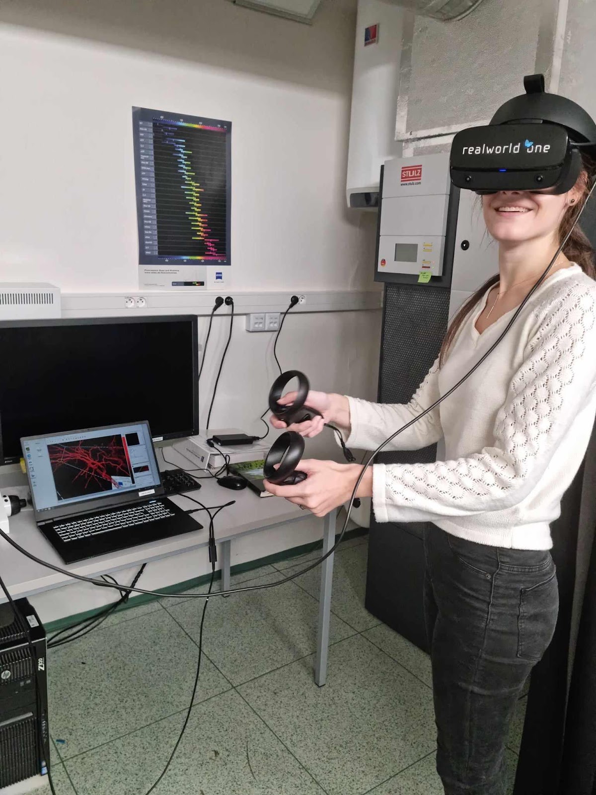
Katerina Kaduchova tests out the Virtual Reality headset at the IEB Plant imaging training course in Prague, October 2023.
This year, the fun revolved around the newly acquired Virtual Reality software and headset. In July, IFIEB invested in an image analysis software Arivis Vision 4D and Arivis Virtual Reality. The idea is to use the Virtual Reality headset to dive into the plant structures, e.g. roots, to see and analyze the structures in 3D/4D from inside. “With the Virtual Reality headset, the image analyst can select and measure a specific deep tissue cell of interest without any segmentation, simply by clicking on it,” explains Katerina M.
“We also use the Virtual Reality headset as an attractive PR tool. It works well, particularly with young students,” she explains. “But we are still exploring the potential of this technology for our work. It is promising, so I’m sure it will find its place in our facility,” she concludes.
From image analysis with Virtual Reality to vertical stage long-term imaging, ultrafast spinning disc, to Airyscan2 with VAHeat, the Multi-modal light microscopy imaging in plant research training course is an excellent opportunity to try out the fabulous range of equipment for plant researchers at IFIEB Prague and get to know the expert staff.
This course is open to external participants, so if you are interested in attending next year, please get in touch!
Picture taken by: Beáta Strejčková (shared by @pecinka_grp on X)
More news from Euro-BioImaging
July 28, 2026
Global BioImaging Advance the Conversation on Sustainable Biomedical Imaging Facilities
How can biomedical imaging facilities remain scientifically excellent while becoming more financially, environmentally, and operationally sustainable? These questions were at the heart of…
Moara Lemos: New insights from Global BioImaging’s Job Shadowing programme
Moara Lemos is a Brazilian researcher and imaging scientist. Her award winning research pushes the boundaries of structural biology, using advanced cryo-electron microscopy…
A Multiplexed Vision: Highlights from the Special Edition Virtual Pub on Spatial Transcriptomics
On June 12th, Euro-BioImaging hosted its latest Special Edition Virtual Pub, bringing together over 200 researchers and technology developers working at the cutting…