
March 26, 2026
Welcome, MAdMiC Node!
The Madrid Advanced Microscopy Center (MAdMiC) is the first Euro-BioImaging Node in Madrid (Spain). It is formed through the collaboration and close work of…

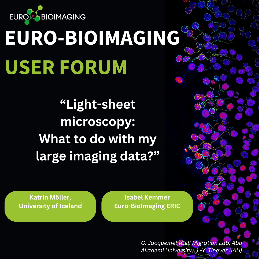
Join us at the Euro-BioImaging User Forum on Image Data, for an afternoon of presentations from Euro-BioImaging users and image data/image analysis experts at our Nodes that will provide a compelling overview of the state-of-the-art in Image Data. At this event, Katrín Möller of the University of Iceland, and Isabel Kemmer, Euro-BioImaging’s Image Data Steward, explore their experience of submitting large light-sheet microscopy datasets to the BioImage Archive.
What: Euro-BioImaging User Forum “Image Data”
When: March 26, 2024, from 14:00-17:00 CEST Where: Online
Abstract
Light-sheet microscopy: what to do with my large imaging data?
Katrín Möller,
University of Iceland
Isabel Kemmer
Euro-BioImaging ERIC
Many of our current biological questions cannot be answered without visualising the object, interaction, or behaviour under investigation. Therefore, microscopy has become an essential tool for many life scientists. While microscopy technologies are rapidly advancing, with increasing speed, resolution, and sensitivity, the amount of data these instruments generate is also steadily growing.
This presents a new challenge for most institutions: how to manage and store their large datasets.
During her doctoral research Katrín Möller used state-of-the-art light-sheet microscopy to investigate, in real-time, the dynamic movements of various intracellular components within microglia, the immune cell of the brain, during their crucial role in clearing apoptotic cells from the developing zebrafish brain.
This research generated several terabytes of data, taking up significant storage space even after careful curation. As publication and a lab transition approached, securing a long-term, open-access repository for this valuable dataset became necessary. Fortunately, the BioImage Archive came to the rescue.
March 26, 2026
The Madrid Advanced Microscopy Center (MAdMiC) is the first Euro-BioImaging Node in Madrid (Spain). It is formed through the collaboration and close work of…
March 26, 2026
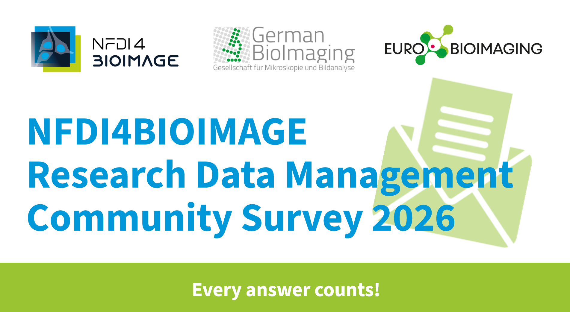
German BioImaging, within its work in the NFDI4BIOIMAGE consortium, and in collaboration with Euro-BioImaging ERIC, has launched a new survey to collect input about…
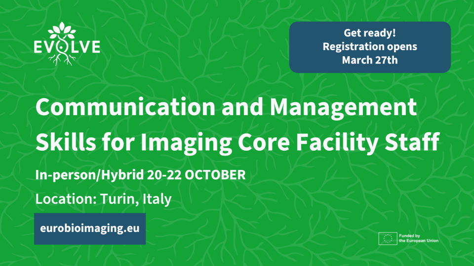
March 25, 2026
Turin, Italy – 20–22 October 2026 Early career professionals working in imaging core facilities will soon have the opportunity to strengthen essential skills beyond…