
March 30, 2026
Euro-BioImaging at the EOSC ESFRI meeting in Milan
Euro-BioImaging was delighted to attend the ESFRI/EOSC Policy Workshop on “EOSC and Research Infrastructures: Opportunities and Strategies” in Milan from March 15-16, represented by…

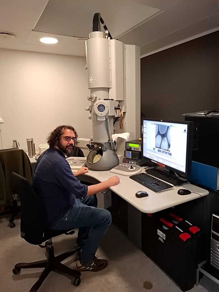
As part of our ongoing series spotlighting the talented scientists working at the Euro-BioImaging Nodes, our Scientific Ambassador Lieke Stockmann sat down with Cristiano di Benedetto, a microscopy specialist at the Core Facility for Integrated Bioimaging (CFIB), part of Danish BioImaging, formerly CFIM, located in Copenhagen. With a background in biology and a career spanning marine biology, glioblastoma research, and advanced electron microscopy, Cristiano brings a wealth of experience and a unique perspective to his role. As CFIB transitions into a new organizational structure under the Center for Core Facilities (CCF), Cristiano finds himself at the intersection of tradition and innovation—both in terms of technology and team dynamics.
Article contributed by Euro-BioImaging Scientific Ambassador, Lieke Stockmann (University of Copenhagen, Denmark)
Cristiano’s journey into microscopy began in Milan, Italy, where he did his PhD in animal biology and used electron microscopy to investigate regeneration in sea urchins. After a postdoc in Saudi Arabia, where he worked on glioblastoma cell cultures and taught electron microscopy, he joined CFIM in Copenhagen in 2017. Initially hired as a lab technician specializing in sample preparation for electron microscopy (EM), Cristiano is now transitioning toward a more imaging-focused role.
"I used to prepare samples from bacteria to human biopsies,” he explains. “Now I’m moving from sample preparation to imaging. I have one foot in each world.
This evolution reflects both personal growth and the broader changes within CFIB, which recently merged with the NMR core facility to form a more integrated bioimaging center. The merger aims to centralize resources and expertise, making it easier for researchers to access imaging technologies across modalities.
Cristiano works primarily with electron microscopy, a field that offers unparalleled resolution and structural detail. He explains the difference between room temperature EM and cryo-EM with clarity and enthusiasm.
Room temperature EM typically involves chemically fixing samples, embedding them in resin, and slicing them into ultra-thin sections—sometimes as thin as 50 nanometers. These sections are then imaged using transmission electron microscopy (TEM), which projects electrons through the sample to create high-resolution images. For surface imaging, a scanning electron microscope (SEM) is employed, allowing researchers to examine larger samples, even small animals. CFIB is equipped with the serial block-face SEMs for 3D reconstruction, and a plasma FIB-SEM that uses focused beams to slice and image samples.
Cryo-EM, on the other hand, preserves samples in a near-native state by flash-freezing them, resulting in vitreous ice that contains no crystals. “Cryo avoids the artifacts introduced by chemical fixation,” Cristiano says. “It’s closer to reality, but much more challenging. You have to maintain a cold chain at -150°C throughout the entire process.”
CFIB offers a diverse range of EM technologies, including serial block-face SEMs, focused ion beam systems, and high-end TEMs like Titan Krios, Glacios, and the versatile Tecnai G2. Some of these instruments support both room temperature and cryo workflows. Cristiano regularly advises users on selecting the most suitable technique based on their scientific needs.
Cristiano’s role is not limited to imaging. He also trains users — from hospital researchers to PhD students — in sample preparation and microscope operation. CFIB offers a tiered support system: full support for beginners, minimum support for intermediate users, and “do-it-yourself” access for experienced researchers.
“I enjoy teaching,” he says. “It’s rewarding to see users become confident and independent. And it’s a good way to stay challenged and avoid routine.”
Despite the technical complexity of EM, Cristiano emphasizes the importance of collaboration and communication. Sometimes he works with light microscopy colleagues to perform correlative light and electron microscopy (CLEM), a technique that combines fluorescence imaging with EM to provide both functional and structural insights.
“CLEM is time-consuming but very satisfying,” he notes. “You need to find the exact same cell imaged with fluorescence and then locate it again in EM. It’s not rocket science, but it takes planning and patience.”
Cristiano’s favorite part of the job? Sitting alone in a dark room with a microscope, radio playing softly in the background.
“It’s relaxing,” he says. “You’re focused, in control, and discovering something new. I also love working with ultra-microtomes—cutting those perfect slices is an art.”
He has a particular fondness for imaging cilia from pediatric nasal biopsies, a task that requires precision and care. “These are kids,” he says. “We want to give the best quality images possible. Sometimes the sample isn’t good enough, and we have to ask for another. But I do my best to avoid that.”
As CFIB grows, Cristiano sees exciting opportunities ahead. He’s particularly interested in mastering new instruments like the Hydra Bio Plasma FIB-SEM and expanding his expertise in cryo-EM and 3D imaging.
“I want to become more of a microscopy specialist,” he says. “Take on more responsibility, teach more, and stay flexible. The future is about adaptability.”
He’s also optimistic about the facility’s direction under the new CCF umbrella. Monthly meetings with other core facilities, including the animal and proteomics units, foster a sense of community and shared purpose.
“There’s a good energy here,” he says. “We have young, dynamic colleagues in light microscopy and experienced hands in EM. It’s a nice mix.”
For young scientists considering a career in microscopy, Cristiano offers thoughtful advice.
“This is a great place to learn,” he says. “You gain technical skills, social skills, and a sense of responsibility. But you’re not doing your own research—you’re supporting others. So you have to ask yourself: what do you really want?
He recommends spending a few years in a facility like CFIB to build a strong CV and gain experience before deciding whether to pursue independent research or move into industry. “For technicians, it’s one of the best jobs you can have,” he adds. “You get to work with amazing instruments and be part of cutting-edge science.
March 30, 2026
Euro-BioImaging was delighted to attend the ESFRI/EOSC Policy Workshop on “EOSC and Research Infrastructures: Opportunities and Strategies” in Milan from March 15-16, represented by…
March 30, 2026
Euro-BioImaging was delighted to attend the Public Awareness & Engagement of Research Infrastructures (PAERI) conference, represented by External Communications Officer, Marianna Childress-Poli. This year’s…

March 26, 2026
The Madrid Advanced Microscopy Center (MAdMiC) is the first Euro-BioImaging Node in Madrid (Spain). It is formed through the collaboration and close work of…