
March 30, 2026
Euro-BioImaging at the EOSC ESFRI meeting in Milan
Euro-BioImaging was delighted to attend the ESFRI/EOSC Policy Workshop on “EOSC and Research Infrastructures: Opportunities and Strategies” in Milan from March 15-16, represented by…

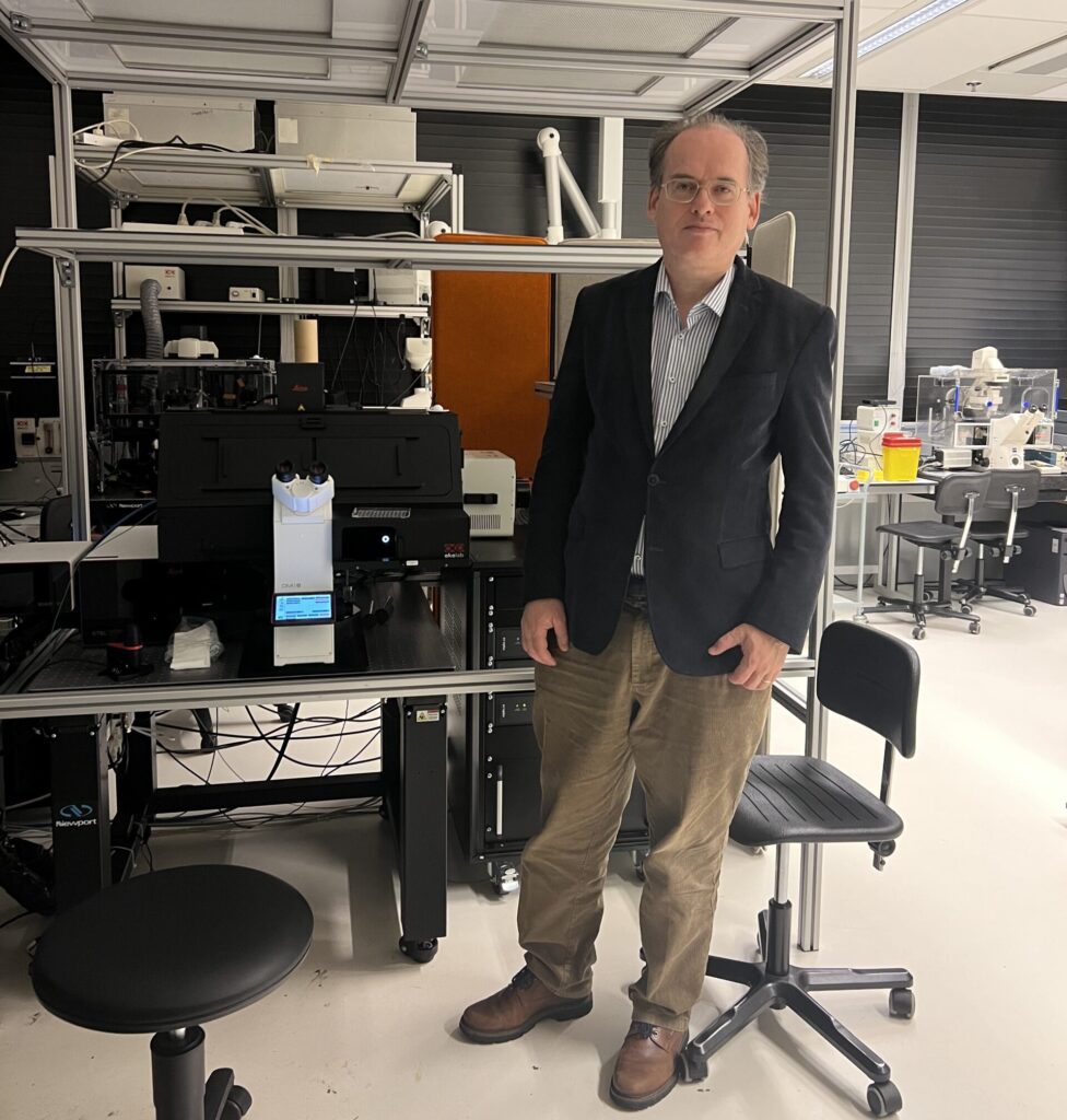
As part of our ongoing Node interview series led by Scientific Ambassadors, Vanessa Coelho-Santos, Group Leader at University of Coimbra, sat down with Professor Dorus Gadella, Director of the Light Microscopy and Imaging Centre (LCAM) in Amsterdam, part of the Dutch LCAM- Functional Imaging Flagship Node within Euro-BioImaging. Join us as we meet Dorus Gadella, who shares insights into his work at LCAM, the evolution of fluorescence imaging technologies, and the collaborative spirit that defines the Amsterdam microscopy landscape. We also explore his scientific journey, the challenges and breakthroughs along the way, and his vision for the future of functional imaging and training the next generation of researchers.
LCAM plays a central role in advancing fluorescence microscopy and functional imaging technologies, driving innovation in the visualisation of dynamic molecular processes within living cells.
LCAM operates as a collaborative infrastructure across three innovative microscopy centres at the Academic Medical Centre (AMC), the Faculty of Science of the University of Amsterdam (FNWI) and the Netherlands Cancer Institute (NKI). This partnership brings together complementary expertise spanning molecular cytology, medical imaging, and cancer research, fostering a comprehensive environment for the development and application of advanced microscopy methods.

Notably, LCAM holds a special place in the history of microscopy in the Netherlands, it was home to one of the world’s first confocal microscopes, marking the beginning of a long tradition of technological excellence and innovation in advanced imaging. Since then, LCAM has continued to expand its infrastructure, offering open access to state-of-the-art microscopy platforms, including confocal and wide-field systems, fluorescence lifetime imaging (FLIM), Förster resonance energy transfer (FRET), fluorescence correlation spectroscopy (FCS), fluorescence recovery after photobleaching (FRAP), stimulated emission depletion (STED), and spectral imaging, among others. Alongside access to high-end instrumentation, LCAM offers technical assistance, user training, specialized courses, and one-on-one methodological support for both local and international researchers.
Professor Gadella and his team are deeply engaged in functional imaging, signal transduction research, and the development of fluorescent proteins and biosensors, fields that are vital for understanding complex cellular behaviors. Under his leadership, LCAM not only serves as a technological hub but also as a vibrant training ground for young scientists, offering advanced microscopy courses and contributing to the broader Dutch and European imaging communities through NL-BioImaging and Euro-BioImaging initiatives.
I have acted as Director since 2010 when we founded LCAM. Before we had the Centre for Advanced Microscopy at Molecular Cytology, UvA, founded in 2003. In June 2011 we started NL-BioImaging AM and LCAM was the nucleation point for organizing the Dutch microscopy landscape and integration into Euro-BioImaging.
The collaboration started in 2009 in view of the Euro-BioImaging developments. LCAM was a formal collaboration between three facilities at three places in Amsterdam: the FNWI (faculty of science), the AMC (academic hospital) and the NKI (Dutch cancer institute). The main reason for collaborating was because the three PIs shared a research agenda on development and application of functional imaging microscopy and because with the combined instrumentation we could engage in larger microscopy courses for our own students and for international students. Benefits of working together included: better visibility, larger microscopy infrastructure, informal contacts with peers. Challenges were mostly linked to travelling across Amsterdam, sometimes admin.
The course we currently run is the Advanced Microscopy course in the Master Biomedical Sciences at the UvA. Currently we have ~ 20 students enrolled in this course. It lasts 1 month, and is intense. We also provide basic microscopy training (1 day courses), a basics in microscopy course (3 days, once/year), advanced microscopy (1x per year) and an international FEBS advanced course (June 2025, which is scheduled every other year). Rest of year several 1:1 training at microscopes. 100 internal users.
Functional imaging, signal transduction, fluorescent protein and -biosensor development are our specialities. We work primarily with confocal, wide field microscopy, and with the more advanced techniques such as FLIM, FRET, FCS, FCCS, FRAP, STED, spectral imaging. We offer access to all these techniques to external visitors.
We work primarily with mammalian cells in culture at our own group. In the institute we provide support also for microbiology, plant and neuro-biology. I enjoy working with multi-well plates with many different biosensor and FP variants and fully automated quantitative imaging.
In terms of sample mounting: plant root hairs for FCS. In terms of sample fluorophores: a new RFP that was chemically instable. It took more than a year to introduce mutations to render it chemically stable.
The contact with enthusiastic young people and the several serendipitous findings we have made in optimization of new fluorescent proteins.
I was interested in biology and how ‘life’ works. So I started studying chemistry since I assumed the secret is in the biomolecules. Biology was quite descriptive at that time. I worked on lipid transport proteins with fluorescent lipid analogs. This triggered my interest in fluorescence spectroscopy, which is exquisitely sensitive and there is no need to separate components and one can continuously monitor kinetics by recording as a function of time. During my PhD I was already doing FRET, synthesis of fluorescent lipids and ns-time resolved spectroscopy. Then I did a postdoc at the MPI Göttingen (Jovin lab). That was a transforming experience as he showed that everything in spectroscopy could also be done using a fluorescence microscope. We did FRET, FLIM spectral imaging etcetera in the early 1990s. That was a pioneering lab. Then I did a postdoc in Wageningen to assemble the first frequency domain FLIM instrument in the Netherlands. This was successful. Then I did another postdoc in Wageningen to apply this technology to plant cell biology, working on plant-microbe interactions. During the end of that time both Mark Hink and Joachim Goedhart were PhD students there. Then I became professor of Molecular Cytology in Amsterdam in 2001 and have been working here since. In time, both Mark Hink and Joachim Goedhart joined the group and both were essential to develop LCAM and functional imaging at our institute. I always had a particular interest in FLIM. The technique has been around since the 1990s, but it was often seen as a too specialistic niche technique. Currently there is a massive revival of FLIM. It has become much more user-friendly and data-analysis is no longer the bottleneck. It is the most quantitative functional imaging technique. It can also sharpen-up images from STED microscopy. We recently acquired a new tau-STED setup in the framework of NL-BioImaging. This utilizes FLIM contrast to enhance super-resolution. We are also busy with novel biosensors that exclusively show lifetime contrast (no other spectroscopic contrast). I see a bright future for FI. So we can image biochemical processes live and quantify the molecular states. It will certainly move in the direction of multiparameter and multiplexing.
I see my own role as quite constant, leading and providing opportunities for young researchers to master, apply and expand these techniques for their own research. I am looking forward to the start of a new consortium Vascular Immunology that has just started with 6 PhD students working in the FNWI and AMC together on one programme. I hope to make a difference there with the team to apply new molecular sensors and FPs to understand the mechanism and issues for migration of white blood cells through the vascular endothelium. In that sense I am living my dream because we are now able to visualize, manipulate and quantify cellular and molecular behavior in intact living cells in an unprecedented way. We live in the golden age of microscopy.
Go for it. Try to learn many different techniques and do not only interact with the software of the microscope. Try to look inside the microscopes, know something about detectors, computers, fluorescence spectroscopy, and probes, and think molecularly as if you would be the fluorescent probe yourself and the microscope is there to find out what you are doing inside the cell.
Every time we organize the FEBS course, when we succeeded to get NL-BioImaging funded, that I am active as member of the Euro-BioImaging board representing NL-BioImaging and that Eric Reits at the LCAM-AUMC is chairing NL-BioImaging.
March 30, 2026
Euro-BioImaging was delighted to attend the ESFRI/EOSC Policy Workshop on “EOSC and Research Infrastructures: Opportunities and Strategies” in Milan from March 15-16, represented by…
March 30, 2026
Euro-BioImaging was delighted to attend the Public Awareness & Engagement of Research Infrastructures (PAERI) conference, represented by External Communications Officer, Marianna Childress-Poli. This year’s…

March 26, 2026
The Madrid Advanced Microscopy Center (MAdMiC) is the first Euro-BioImaging Node in Madrid (Spain). It is formed through the collaboration and close work of…