
March 26, 2026
Welcome, MAdMiC Node!
The Madrid Advanced Microscopy Center (MAdMiC) is the first Euro-BioImaging Node in Madrid (Spain). It is formed through the collaboration and close work of…

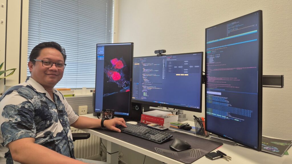
As part of our ongoing series spotlighting the talented scientists working at the Euro-BioImaging Nodes, we sat down with Junel Solis, an expert in image data analysis and data management based in Turku, Finland. Junel’s work supports researchers across disciplines, helping them make sense of increasingly complex imaging data. We asked Junel about his role, the diverse projects he encounters, and what keeps him motivated in this fast-changing field.
Article contributed by Euro-BioImaging Scientific Ambassador Pablo Suarez-Cortes (Max Planck Institute for Infection Biology, Berlin)
Could you introduce yourself? What’s your role, and how long have you been here?
My name is Junel Solis. I’m the Head Image Data Analyst at the Finnish Advanced Microscopy Node of EuroBioImaging, and I’ve been in this position for almost five years.
What is your expertise and what do you do at the Node?
My main focus is on image data management and image data analysis. We provide image data analysis as a service—it’s basically open access for anyone needing help with their imaging datasets. Researchers come to us with their data, tell us a bit about their research and what they hope to achieve, and then we work out how best to process, analyse, and manage their data.
Sometimes, that means using established tools; other times, it involves building something customized and reproducible to address a unique challenge. An increasingly important part of our work is data management: figuring out where and how to store data, in which formats, and how to visualize and share it efficiently.
What kind of samples and projects do you typically work with?
We work predominantly with biological imaging data—especially confocal microscopy images, which is where the unit started. But, in recent years, our scope has expanded far beyond classical cell biology.
Here are some of the more unusual projects we’ve encountered:
It’s always exciting when something completely new comes our way. We just have to read up on the topic and figure out how to extract useful information from unfamiliar data.
Despite all this variety, our "bread and butter" remains biological microscopy—especially light and electron microscopy—but we love branching out when we can.
What kinds of challenges do you face in your work?
The biggest challenge is often the unknown. Some samples are familiar—say, standard cell images—but sometimes we get data that’s completely outside our usual expertise. As an analyst, you can look at an image and spot certain features. But unless you have background knowledge—say, knowing what a "blob" actually signifies in a cell—you risk making incorrect assumptions.
That’s why it’s crucial to interact closely with the researcher supplying the data. Their input is essential in the analysis loop, especially when it comes to interpreting the biological significance of the results.
Another huge challenge nowadays is handling the scale of the data. Image files from modern microscopes can be staggeringly large—sometimes a single image is already several gigabytes. Storage capacity isn’t the main problem—it’s being able to view, process, analyse, and share these massive datasets quickly and reproducibly. As images scale from simple 2D snapshots to vast, multi-channel 3D datasets or time series, the technical challenges multiply. You need efficient strategies to process and store such data without overwhelming your computer.
What do you like most about your job?
Above all, the challenge. There’s always a new puzzle to solve—whether that’s coming up with faster analysis, finding a way to process a massive dataset, or inventing tools to handle data we’ve never seen before. Sometimes I find myself thinking about an analysis problem the night before, and I’m excited to try new solutions the next morning.
My main focus is on image data management and image data analysis. We provide image data analysis as a service—it’s basically open access for anyone needing help with their imaging datasets. Researchers come to us with their data, tell us a bit about their research and what they hope to achieve, and then we work out how best to process, analyse, and manage their data.
- Junel Solis, Finnish Advanced Microscopy Node
But it’s also about enabling others: whether it’s cell biologists, cancer researchers, agronomists, or marine ecologists, I feel proud knowing our work helps so many different areas of science move forward.
How did you get here—what was your career path like?
Ironically, I didn’t set out to become an image data analyst! As a teenager, I played around with popular image processing programs; that gave me a sense of how digital images work—looking at pixels up closely, manipulating colours and features.
My background is in medicine—my parents are both surgeons, and I grew up in a very remote area of West Africa where they worked in a small village hospital with very few resources. That experience led me to study medicine myself. After I became a licensed doctor, though, I realized I wanted to contribute in a different way.
I moved to Finland and enrolled in a master’s program in biomedical imaging. That’s where my focus switched toward image analysis. I didn’t have a strong background in programming initially, so I had to learn a lot of those skills myself—from both formal courses and self-study.
How do you see your— and the field’s—future?
The field is evolving rapidly: data volumes and complexity are increasing, and the range of application areas continues to expand. There’s a huge need for people who can bridge the gap between technical solutions and real-world scientific problems. For upcoming scientists, I’d encourage learning both the domain science (biology, for example) and strong computational skills. Being able to communicate clearly with researchers from different backgrounds is vital.
For myself, I’d like to continue growing in this multidisciplinary environment—helping researchers tackle whatever new challenges imaging science throws our way.
Junel’s story is just one example of the expertise and dedication you’ll find in EuroBioImaging’s pan-European network. Stay tuned for more profiles from our Nodes community!
March 26, 2026
The Madrid Advanced Microscopy Center (MAdMiC) is the first Euro-BioImaging Node in Madrid (Spain). It is formed through the collaboration and close work of…
March 26, 2026
German BioImaging, within its work in the NFDI4BIOIMAGE consortium, and in collaboration with Euro-BioImaging ERIC, has launched a new survey to collect input about…
March 25, 2026
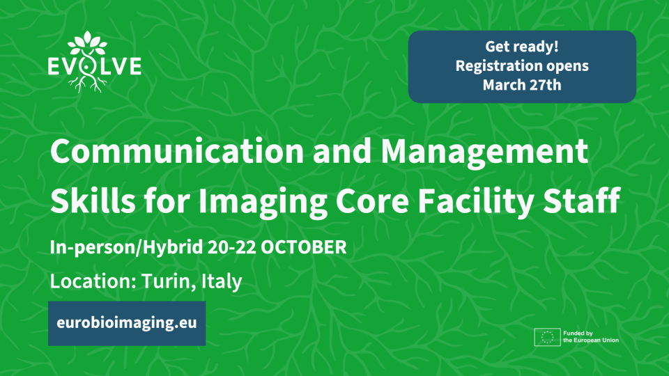
Turin, Italy – 20–22 October 2026 Early career professionals working in imaging core facilities will soon have the opportunity to strengthen essential skills beyond…