
March 30, 2026
Euro-BioImaging at the EOSC ESFRI meeting in Milan
Euro-BioImaging was delighted to attend the ESFRI/EOSC Policy Workshop on “EOSC and Research Infrastructures: Opportunities and Strategies” in Milan from March 15-16, represented by…

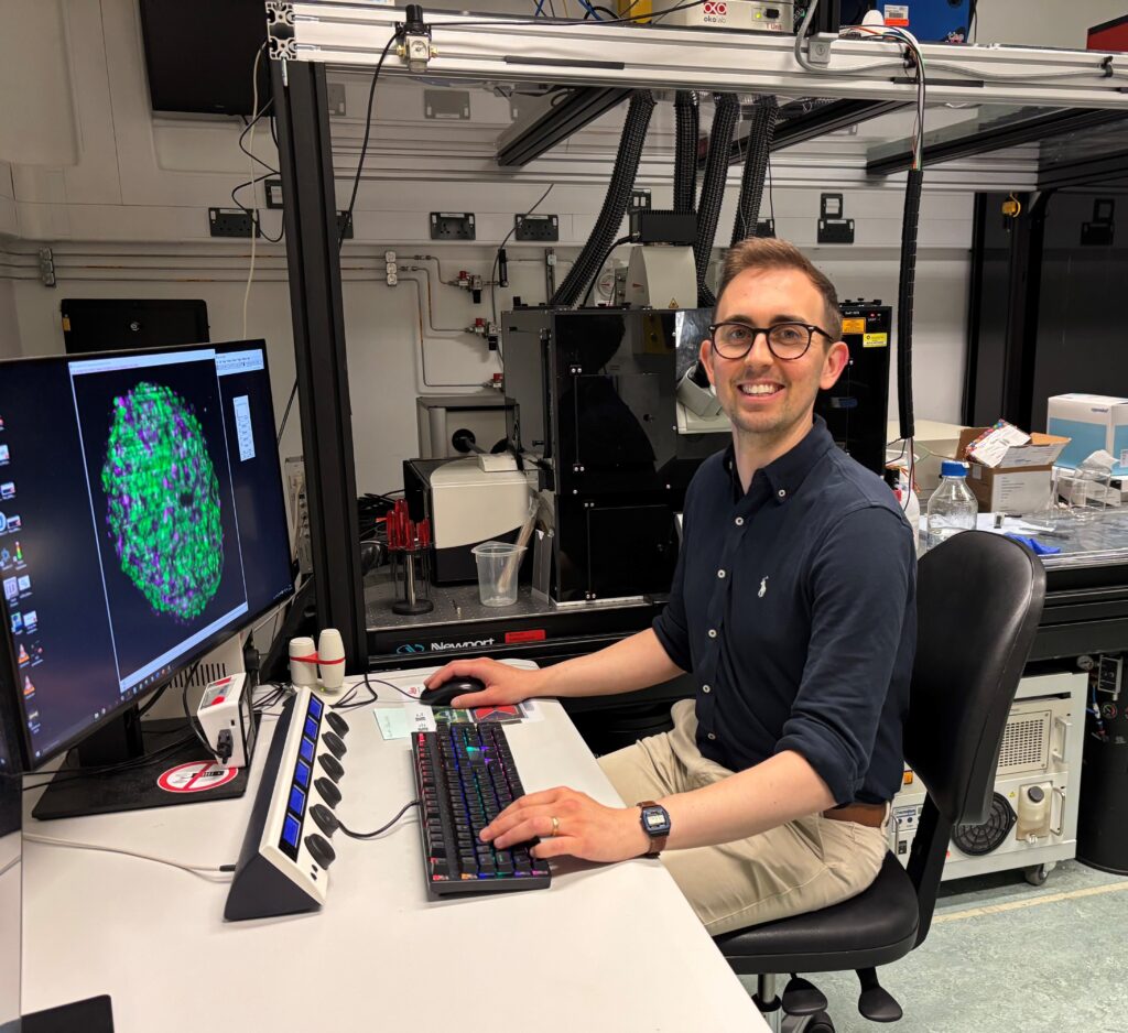
As part of our ongoing series spotlighting the talented scientists working at the Euro-BioImaging Nodes, we sat down with Robert Lees, an expert in multiphoton and lightsheet microscopy based at the Octopus Facility in Harwell, part of Euro-BioImaging’s UK Node. As a Link Scientist, Robert plays a central role in supporting visiting users from across the biological and physical sciences. With a background in cell biology and microscopy, he works closely with researchers to help design experiments, troubleshoot imaging workflows, and develop tailored solutions for complex samples. We asked Robert to tell us about his role and the diverse projects he encounters.
Article contributed by Euro-BioImaging Scientific Ambassador Virginia Silió (University College London, UK)
Located at the heart of the UK’s national research infrastructure, the Octopus (Optics Clustered to OutPut Unique Solutions) team at the Central Laser Facility collaborate with users to design imaging experiments utilising advanced and bespoke imaging technologies. The facility is part of the Science and Technology Facilities Council (STFC), with a suite of advanced fluorescence and electron microscopy techniques—ranging from fluorescence super-resolution and light sheet to correlative and multi-modal pipelines.
Rob’s work is grounded in fluorescence-based imaging techniques, with a particular focus on light sheet fluorescence microscopy. He has developed substantial experience in optimising light sheet workflows for a wide range of samples from live spheroids and organoids, to fixed and cleared whole mouse organs, human skin models and even human organ biopsies.
“I’ve worked with users to image whole mouse eyes, showing the cornea all the way through to the optic nerve,” he explains. “That involved quite a lot of sample handling troubleshooting, making sure the tissue was mounted stably, and figuring out how to scan through the whole thing without obscuring the most important parts.”
Much of the cleared tissue work involves extensive trial and error around sample preparation and labelling to achieve optical clarity and uniform signal. Rob notes that even small differences in tissue processing can affect the final image quality.
“I spent a lot of time trying to optimise clearing, and one of the challenges is that the protocols are often very particular to a specific tissue or fluorophore. That’s something we really need more community support for. A dream for the future would be to create a tissue clearing wiki where you could search for answers to your questions from real experiences, rather than relying on publications with no feedback. This is why I joined and now help to organise the international Slack group for light sheet microscopy, Wildsheet, with over 400 members worldwide.”
While his expertise lies in imaging, Rob’s role spans the full experimental pipeline. From advising on sample preparation, to setting up imaging protocols and managing large datasets, his aim is always to help users get the most out of their time on-site.
“I don’t build microscopes, but I help get the whole pipeline working for our users,” he says. “We’re often trying to make everything run smoothly—whether that’s prepping samples, imaging them, or getting the data stitched and ready to analyse. We collaborate with users before and after their visit too, teaching them about microscopy and image analysis.”
Working at Octopus means engaging with researchers from many different disciplines, often on short timescales.
“A typical visit might be up to two weeks,” he says. “People usually come with samples and a clear idea of what they want to do, but they don’t always have experience with the equipment. My job is to help them figure out what’s realistic, adapt their protocol, and work around any challenges, and get a workable solution ahead of their access time.”
Rob came to the Octopus Facility in 2022 after a background in cellular neurobiology and systems neuroscience. In his PhD at the University of Bristol, he used live imaging and electron microscopy to study synapse formation in mouse neurons in vitro and in vivo. He later moved into postdoctoral work at the University of Oxford, where he helped set up microscopy systems in a new research group.
“That’s where I really got into facilities work,” he reflects. “I was spending more time helping other people use microscopes and get the most out of the systems, and less time asking questions and doing my own research—but I found I actually enjoyed that change.”
That experience led him to apply for his current role, where he now works as part of a multi-skilled team supporting the UK’s national imaging capability.
Many of the projects Rob supports require adapting standard protocols to suit the facility’s bespoke systems. One such example involved imaging mouse eyes stained for retinal ganglion cells—an effort that required careful balancing of labelling intensity, clearing transparency, and mounting stability.
“It’s definitely one of the more complex pieces of work I’ve done here,” he says. “From preparing the sample to acquiring and stitching the data. After many months of optimisations it was really satisfying to see that come together.”
He works with the Wildsheet Slack community to help build shared resources and communicate best practices around sample preparation and imaging workflows.
“We’re hoping to develop more standardised ways to share that kind of knowledge—whether it’s sample mounting tips or clearing protocols that actually work.”
Looking ahead, the team are in the process of shaping Octopus’ future direction—both in terms of instrumentation and how it positions itself within the UK bioimaging landscape.
“We’re a relatively large facility, and the way we operate is complex—we mainly work with external users. That comes with challenges to make sure we’re offering something unique and impactful”, he says. “One of my hopes is that we better integrate with the wider BioimagingUK network. Octopus is not trying to duplicate what other people do—we’re here to augment the existing infrastructure, to be a hub that complements other facilities”
“Something we’re thinking a lot about is: how do we complement other facilities?” he says. “We don’t want to replicate what other sites are already doing. Our strength is in offering more in-depth, collaborative and specialist support—somewhere users come once they’ve done the groundwork and want to push things further.”
The facility also sees growing interest from industrial users, which brings new demands around reproducibility, documentation, and long-term support.
“Those users often need more structured workflows—and we’re learning how to support that as well.”
As an early-career technical specialist, Rob is also engaged with wider conversations around research infrastructure and technician careers.
“There’s definitely more support now for technical roles, especially with things like the Technician Commitment,” he says. “But we still need clearer paths for progression—especially in facilities where the work is so collaborative and user-focused.”
His own career continues to evolve, and he sees his future in facility management or technical operations management of some sort.
“What I enjoy most is solving problems and helping other people get the data they need by fitting all the pieces together as smoothly as possible. That’s what keeps this job exciting.”
With a collaborative mindset and deep practical expertise, Dr. Robert Lees continues to play a vital role in shaping user-focused innovation within the Octopus Facility—bridging biological research and cutting-edge technology in ways that have lasting impact.
March 30, 2026
Euro-BioImaging was delighted to attend the ESFRI/EOSC Policy Workshop on “EOSC and Research Infrastructures: Opportunities and Strategies” in Milan from March 15-16, represented by…
March 30, 2026
Euro-BioImaging was delighted to attend the Public Awareness & Engagement of Research Infrastructures (PAERI) conference, represented by External Communications Officer, Marianna Childress-Poli. This year’s…

March 26, 2026
The Madrid Advanced Microscopy Center (MAdMiC) is the first Euro-BioImaging Node in Madrid (Spain). It is formed through the collaboration and close work of…