
March 26, 2026
Welcome, MAdMiC Node!
The Madrid Advanced Microscopy Center (MAdMiC) is the first Euro-BioImaging Node in Madrid (Spain). It is formed through the collaboration and close work of…

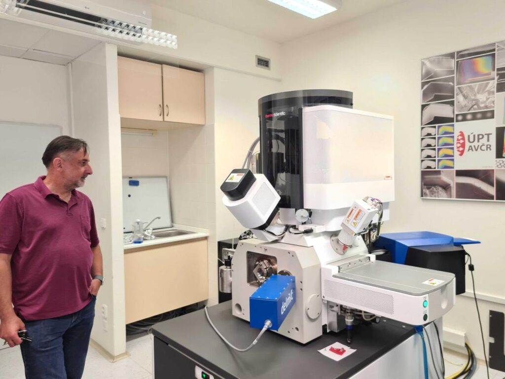
In 2024, the Brno Node brought new facilities into the Node, further expanding its service offer and technology remit. Following the CTLS meeting in Brno, Johanna Bischof, Head of Bio-Hub operations, met with representatives from all Brno Node facilities and visited several of the new facilities within the Institute of Scientific Instrumentation at Masaryk University.
Preclinical imaging facility at the Institute of Scientific Instrumentation:
This facility is a fully open-access preclinical imaging facility, including with a highly professional veterinary setup that allows animals from external institutions to be brought to the facility and hosted there for the duration of imaging experiments. The facility provides 9.4T µMRI wtith a specific focus on functional imaging, specifically perfusion imaging.
“The facilities are all very impressive in their technology offer and expertise. The Brno Node is impressively comprehensive in its technology offer - from Electron and Light Microscopy, to preclinical and clinical imaging and data services. Experts in all these different technology areas collaborate closely and provide support for research in a diverse array of subject areas.”
- - Johanna Bischof, Head of Bio-Hub Operations
Due to the institution’s focus on and available expertise in technology development, the facility experts surrounding Radovan Jirik, Head of the facility, can adapt the imaging workflows beyond what is preset in the instruments by programming the acquisition parameters for functional imaging and reaching increased resolution.
Their application focus areas are on animal models for Parkinson’s disease, depression, ageing, and autism, as well as on collaborations for drug testing with the Institute of Pharmacology.
Center for Biomedical Image Analysis
The CBIA provides advanced image analysis services for both biological and biomedical imaging data. Their experts focus on complex analysis problems, such as developing new software tools to tackle complex and large data analysis problems.
A big focus is on multimodal image analysis and the opportunities provided by combining different data sources, including image data and beyond, in machine-learning models to improve their performance on complex tasks.
They work closely with other facilities within Czech BioImaging to support their data analysis and provide analysis as a standalone service. Additionally, the CBIA team is very involved in the Board for benchmarks and challenges in medical image analysis, which oversees the quality and prioritisation of the global benchmarking initiatives and Challenges.
Raman Imaging facility at Institute of Scientific Instrumentation:
The small and specialised facility focusses on direct Raman Imaging and combining Raman imaging systems with other modalities, such as a combined Raman-Optical tweezer system and developing microfluidics platforms for the Raman platform.
Their application focus lies on microbial samples. Here, a fascinating and impactful potential application of Raman imaging is to gather the Raman profiles of clinical bacterial isolates to allow for a later quick and easy detection of different bacteria strains in blood samples. Such a detection has potential applications in quick testing and prescription of suitable antibiotics against antibiotics resistent strains.
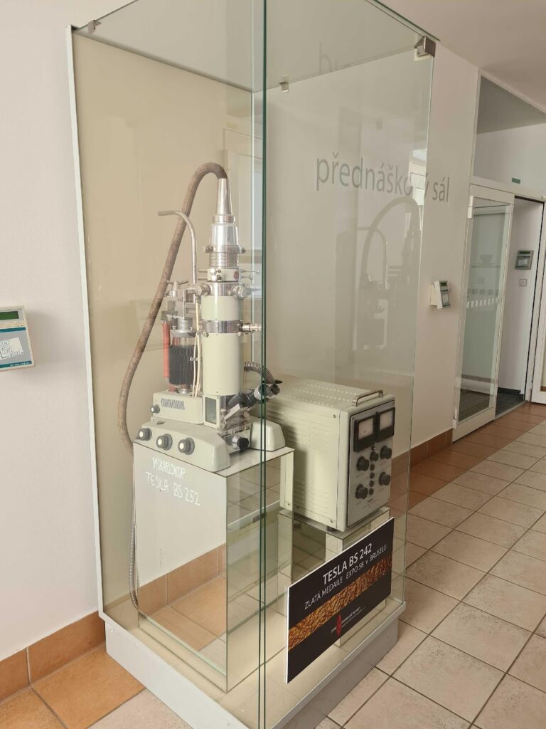
Electron Microscopy at Institute of Scientific Instrumentation:
The EM facility at ISI is a large facility with a broad instrument park and a focus on developing new EM tools and improving EM systems. Due to their expertise in scientific instrument development, they take apart and adapt existing EM systems and develop their own platforms and tools to expand the capabilities of the instruments.
This builds on the history of Brno and the ISI as the cradle of Electron Microscopy.
Their developments cover everything from specialised sample holders, combining Raman Imaging and EM, and a special focus on super-cryo EM with ultralow temperatures below what is achievable with liquid Nitrogen cooling.
The faciliy is very actively collaborating with the local EM companies in Brno on new technical development, specialised applications and training.
All these systems and expertise – as well as the expertise in cellular and label-free imaging and CELLIM and Biophotonics core facilities, and on human MRI imaging at MAFIL – are available for Euro-BioImaging users in open access.
March 26, 2026
The Madrid Advanced Microscopy Center (MAdMiC) is the first Euro-BioImaging Node in Madrid (Spain). It is formed through the collaboration and close work of…
March 26, 2026
German BioImaging, within its work in the NFDI4BIOIMAGE consortium, and in collaboration with Euro-BioImaging ERIC, has launched a new survey to collect input about…
March 25, 2026
Turin, Italy – 20–22 October 2026 Early career professionals working in imaging core facilities will soon have the opportunity to strengthen essential skills beyond…