
July 9, 2026
Euro-BioImaging Visits the NORMOLIM Node at the 2026 180°N Conference
In April 2026, the Med-Hub Head of Operations Alessandra Viale attended the 2026 180°N Conference, held at the beautiful Nye Hjorten Teater…

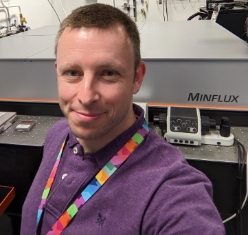
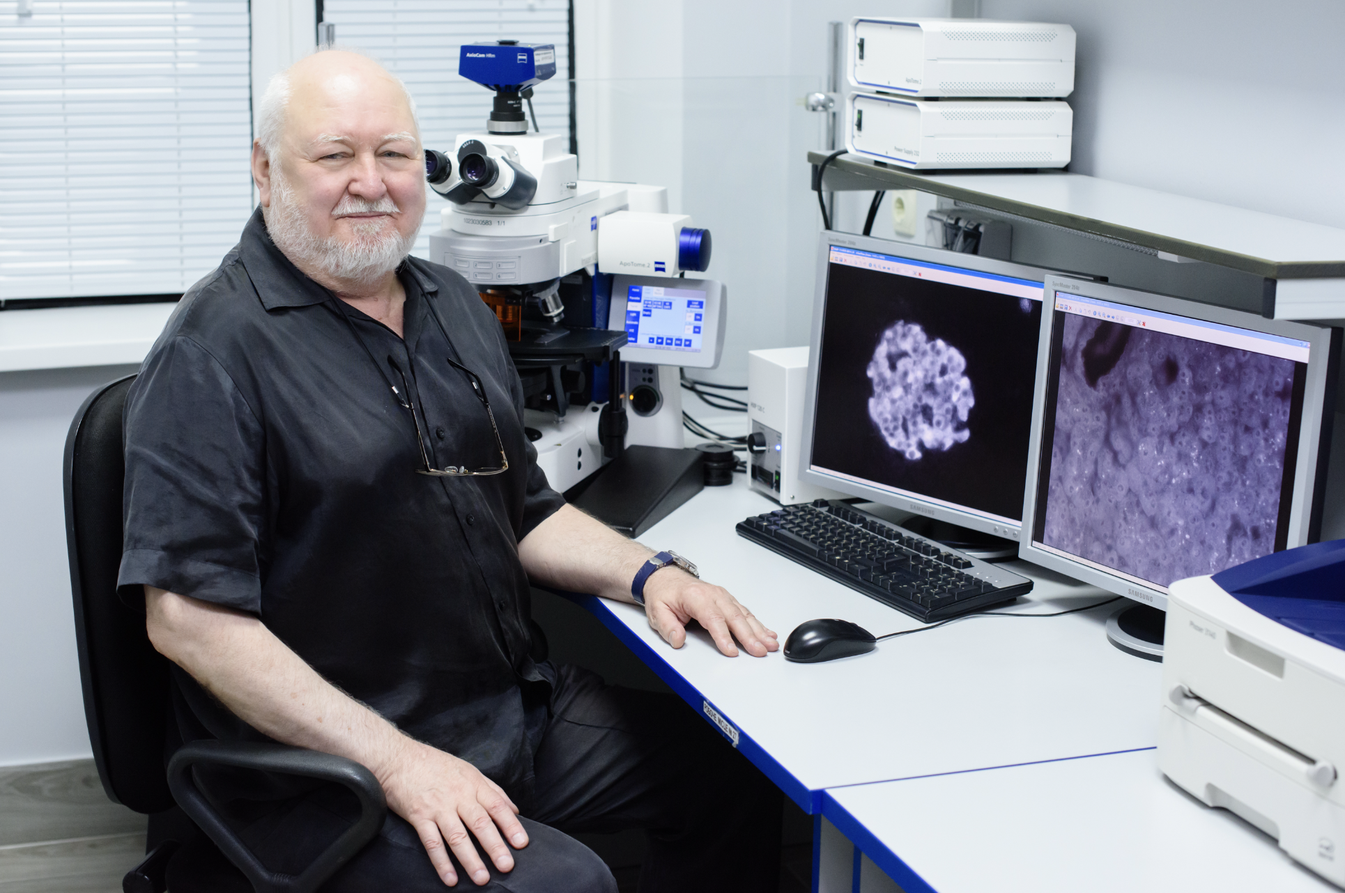
Want to see objects that are 5 nanometers apart? Need to track objects moving in live cells with high temporal resolution? Then why not give MINFLUX a try? Two Euro-BioImaging Nodes are currently offering MINFLUX in open access as part of our Proof-of-Concept study. We spoke to Christopher Tynan, a staff scientist at Central Laser Facility - Octopus Cluster. His facility, which is part of the Euro-BioImaging UK Node , is now accepting applications for MINFLUX projects from external users.
Read more about this technology and what it can be used for in the interview below.
In a nutshell, how does MINFLUX work?
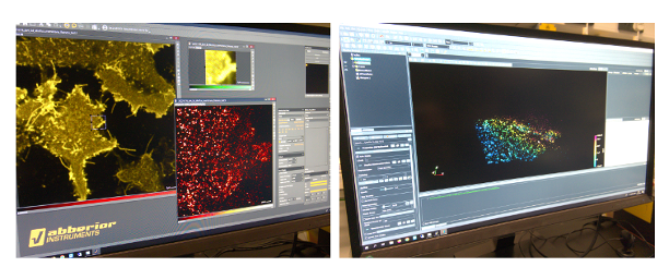
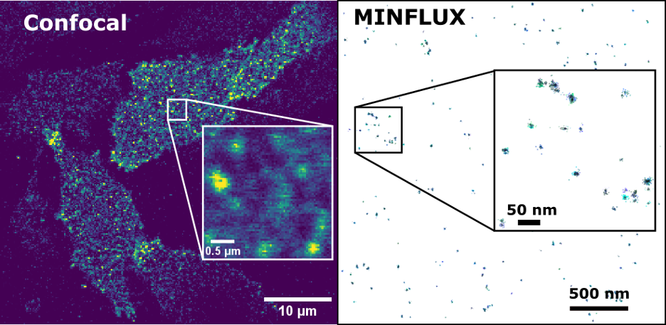
MINFLUX relies on being able to locate individual fluorescent molecules, one at a time over and over again to form an image. Unlike PALM and STORM, where a subset of fluorescent molecules is captured across the whole field of view onto a camera, with MINFLUX, we use a donut-shaped scanning beam that triangulates the emitters one at a time. Each position can be measured very fast, with more precision than can be achieved with a camera while needing to collect far fewer fluorescence photons. The technique gets its name from the phrase ‘minimal emission fluxes’. Nonetheless, it takes a similar amount of time to PALM and STORM to acquire a whole image.
With MINFLUX, we can distinguish dye locations that are around 5 nanometers apart when used for high precision localization. And it is non-destructive because the high sensitivity of detection allows us to use only low amounts of light for the localisation.
What is MINFLUX used for?
This technique is perfect for looking at protein organisation, and protein complexes in cells. Both 2D and 3D imaging is possible. MINFLUX is clearly interesting for people studying cellular structural biology or biological processes at the molecular level, either looking to observe previously unresolvable details in cell structures or at receptor clustering with a higher resolution. For example, people working in neuroscience might want to look at receptors clustering in synapses, or you might want to investigate the organisation of calcium channels in muscle cells. The possibilities are endless because with MINFLUX you can look at cellular processes on the level of individual proteins inside cells.
Generally, any experiment STORM can do, MINFLUX can do with 10x the precision.
Another important application of MINFLUX is object tracking. If you’re willing to trade in a bit of localisation precision, MINFLUX makes it possible to track objects that are moving in live cells with very high temporal resolution. This can be very useful for biological experiments, to follow things that move very fast like lipids or have complicated dynamic properties like membrane proteins. With MINFLUX, you can get a really detailed recording of their movements that you wouldn’t be able to see with camera-based systems. With camera based tracking, the smallest time between measurements is 10 milliseconds. Even without pushing for the highest temporal resolution, with MINFLUX this time step is 0.65 milliseconds on average.
How do you prepare the samples?
What’s key with MINFLUX localisation samples is that they need to be very still. For high-precision localisation, only chemically fixed samples are accepted. In addition gold fiducial beads will have to be added so that an active stabilization system can maintain the sample position. We can obtain less than a nanometer variation in sample position when it’s working right. But with MINFLUX, it’s still really hard to predict how easy it’s going to be to get an experiment working. Some samples require more trouble-shooting, for example, if the sample is too densely labelled. And of course, only a few fluorescent dyes work really well with MINFLUX and ideally we need to keep the dye as close to the target as possible by using a small tag. That all means that it may take us a few days to optimise samples and is also the reason why we let potential users come to our facility before they even apply for access. That way we can assess the level of challenge and demonstrate that the experiment could work in practice.
Tell us a bit more about a specific project that was done in your facility using this technology? What scientific questions were you addressing?
We’ve been doing a lot of work with membrane proteins (most of this work is still unpublished), because MINFLUX lets us see some clusters and complexes that can’t be resolved by STORM.
We published a paper in September 2022, in which we first performed camera based single particle tracking and backed up these results with high precision MINFLUX localisation. Although we were tracking proteins moving together, our user (and a reviewer) also wanted to know if they were close enough to be interacting. We had predicted that our dye labels would be slightly too far apart for other proximity tests such as FRET, so we used MINFLUX to show that dye pairs attached to different receptors in this study were around 10 nm apart.
What other services do you provide in your facility that would be useful in combination with this type of imaging?
There are a range of complementary fluorescence microscopy techniques that users can also apply to use at our facility. Furthermore, we are part of the Science and Technology Facilities Council’s Central Laser Facility which includes the ultra-fast spectroscopy facility ULTRA, and we are situated on the same site as the ISIS neutron source and Diamond Light Source. Collaborative projects across our facilities are highly encouraged.
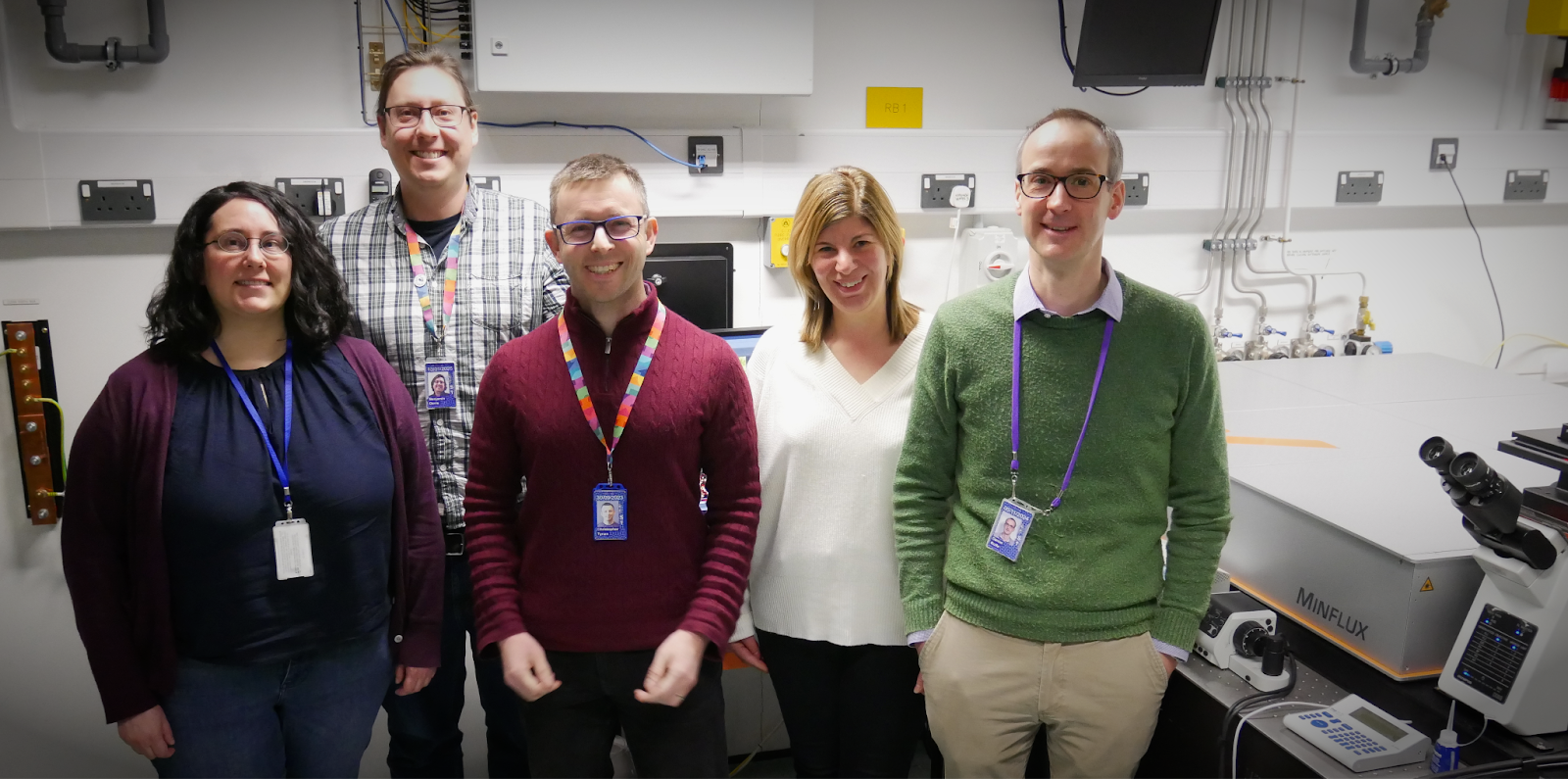
Another reason to work with us is that we have a fully interdisciplinary support team. For example, my background is physics, but we also have cell biologists, biochemists and a data exploitation team who all work with the users. If the project requires it, we’re able to support the user from the planning stage, through sample preparation, with using the microscopes, and then the data analysis. The data analysis support is particularly important for MINFLUX, to translate the localisation information collected into images or quantitatively analyse the distribution of fluorescent dye positions.
We have access to mammalian and insect cell culture, and bacterial culture facilities. Usually people come and prepare samples with us and join us on the microscope to fully learn the technique, including analysis tools. We can also help develop new analysis to extend existing algorithms or address specific spatial analysis questions. It’s a fully collaborative process, from the start to the end of a user’s project.
How to apply:
MINFLUX is part of the Euro-BioImaging Proof-of-Concept study. The Proof-of-Concept study makes it possible to introduce exciting, new imaging technologies to our portfolio that were previously unavailable via our network. We are currently accepting applications to use these technologies at participating Nodes as part of the Proof-of-Concept study. Be part of this study - and contribute to community-wide continuous technological innovation!
All scientists, regardless of their affiliation, area of expertise or field of activity can benefit from Euro-BioImaging’s pan-European open access services.Potential users of these new technologies are encouraged to submit project proposals via our website. To do so, you can log in to access our application platform, choose the technology you want to use and the facility you wish to visit, then submit your proposal. All applications will be processed by the Euro-BioImaging Hub. As usual, users will benefit from advice and guidance by technical experts working at the Nodes, training opportunities, and data management services.
For more information: info@eurobioimaging.eu
July 9, 2026
In April 2026, the Med-Hub Head of Operations Alessandra Viale attended the 2026 180°N Conference, held at the beautiful Nye Hjorten Teater…
July 7, 2026
Armed conflicts generate long-lasting environmental contamination that extends well beyond the duration of military operations. The release of heavy metals such as Arsenic,…
July 6, 2026
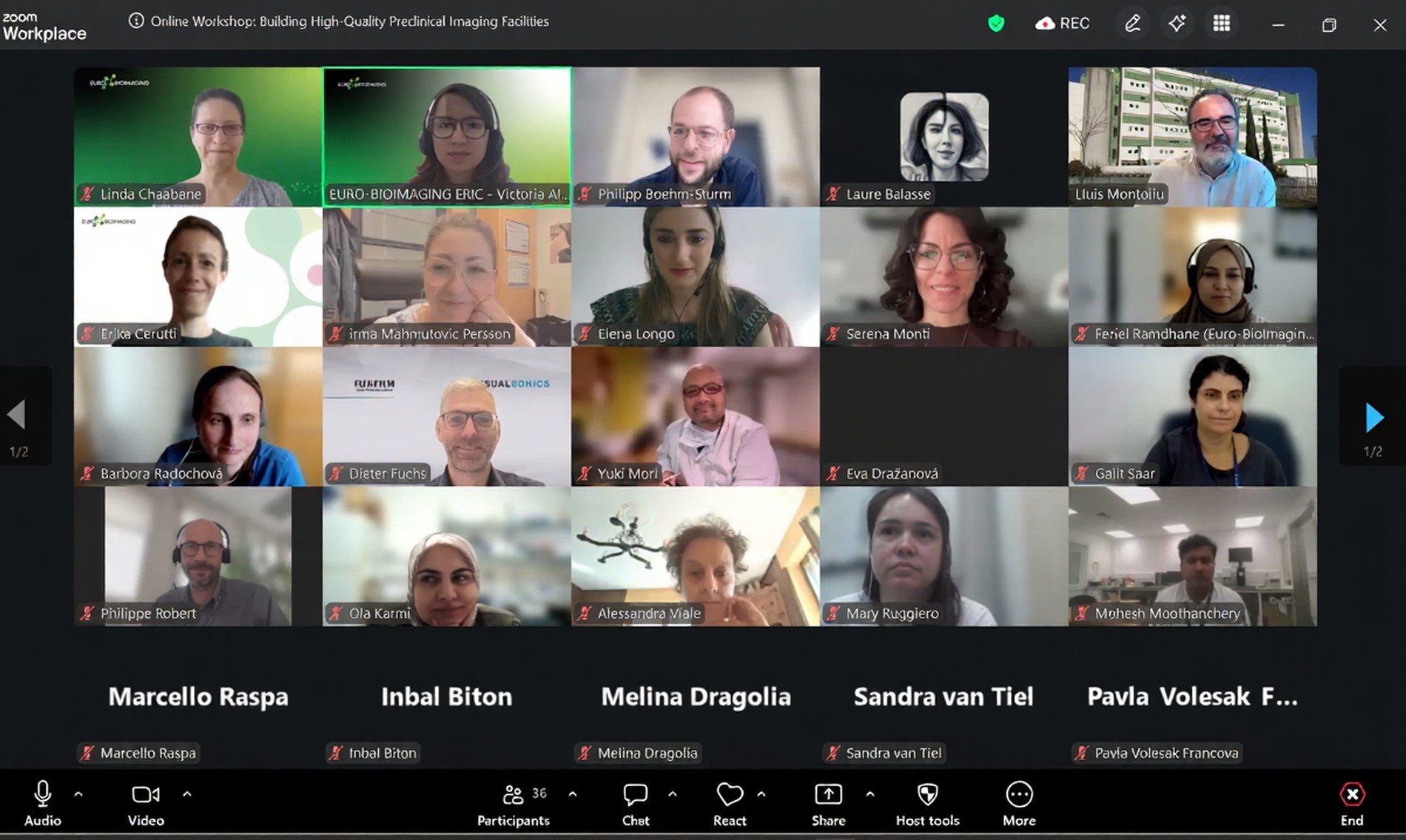
On 2 July 2026, Euro-BioImaging hosted the online EVOLVE workshop “Building High-Quality Preclinical Imaging Facilities”, bringing together approximately 40 imaging facility staff and…