Dr. Alena Salašová is an Assistant Professor at the Department of Biomedicine, Aarhus University (AU). Her work focuses on neurodevelopment, in particular, understanding the cell and molecular mechanisms behind it. With support from the staff at the CEITEC MU CELLIM light microscopy facility, part of our Advanced Light Microscopy and Medical Imaging Node in Brno, Czech Republic, she and her team used light-sheet microscopy to image entire mouse embryos, capturing all developing motor neurons in time and space. Thanks to this amazing imaging work, she, together with the team of Prof. Anders Nykjaer (AU) were able to closely observe the SorCS2 receptor and identify its role in the spinal cord development, the topic of their recent publication in Cell Reports. To learn more about this outstanding Euro-BioImaging user project, we spoke to Alena Salašová and Milan Ešner, the Head of the CEITEC MU light microscopy facility - CELLIM.
“We’ve discovered a new mechanism that tells us about how motor neurons develop, how they make decisions and how they connect with muscles. This could be relevant across different neurological diseases or for spinal cord injury,” explains Alena Salašová enthusiastically. “Looking back, it’s amazing we were able to do this - using an imaging technology like light-sheet microscopy that we did not have access to in our home institution and without any prior know-how.”
From mission impossible to mission accomplished
Indeed, what started out as a very challenging task, got transformed into one of the most beautiful publications of Alena’s career, thanks to the help of Milan Ešner and Jan Křivánek’s teams. “When I started this project,” Alena says, “I knew I had to do 3D reconstructions of the entire mouse embryo in order to revise a paper that was stuck in review. We had to do the experiment from scratch with a very tight deadline - a demanding outset.”
Thanks to a complex network of professional relationships with her collaborators, Alena contacted Milan Ešner, the head of the CELLIM facility in Brno, to accomplish her mission. The CELLIM facility is part of the Euro-BioImaging Advanced Light Microscopy & Medical Imaging Node in Brno, which provides open access to a wide range of imaging technologies and expertise to all scientists, including training scientists in biological and medical imaging techniques and data analysis.
“My alumni student Petra Kompanikova from the Masaryk University once recommended that I should contact Milan at Euro-BioImaging when in need of light-sheet microscopy. I really relied on this personal network since I wasn’t able to travel to the Czech Republic myself. I feel very lucky to have great colleagues there who could help me.”
Alena contacted an old friend, Dr. Jan Křivánek, who heads a lab at the Department of Histology and Embryology, Faculty of Medicine, Masaryk University, and who has proficient experience with the relevant methods. He and his talented PhD student Josef Lavický offered to make the samples transparent, and image them on the light-sheet microscope under CELLIM supervision.
The advantages of light-sheet microscopy
“Light-sheet microscopy allows you to look at big objects in 3-dimensional structure with single cell resolution. But it’s a very complex modality. The sample preparation using the clearing method is crucial to succeed,” says Milan.
Alena agrees. “That’s why Jan and Milan’s teams expertise was so valuable. I had never done light-sheet before and I didn’t have a good clearing protocol. Milan and Jan already had an established pipeline that would allow us to see the single neuronal fibres. And that was a game-changer for me. The light-sheet imaging we did at CELLIM allowed us to look at all the nerves in the whole mouse embryo, something we couldn’t do before.”
During the collaboration, Alena learned a lot about light-sheet microscopy, even from afar. Recently, her department purchased its own light-sheet microscope, so they will be able to apply the expertise gained in the Euro-BioImaging user project and do experiments on their own in the future.
The challenge of data handling and image processing
But the best part was that Alena could do all of the image analysis from Denmark, by connecting to the CEITEC CELLIM Facility’s Virtual Desktop.
“In so many ways, the expertise of Milan’s staff greatly advanced our project,” continues Alena. “After microscopy, we had to analyze the samples. In 3D, that’s anything but trivial.” An expert from Milan’s team trained Josef and Alena to use the latest version of the IMARIS software for the 3D reconstructions and analysis. “
“Moreover, with light-sheet microscopy, you generate huge data sets. We did not want and actually were not able to transfer them in a reasonable timeframe. Especially since the infrastructure for data processing was not yet sufficient at our department,” says Alena.
“We set up a new complete data workflow for Alena,” We were lucky that at the time Biological data management core facility of Ceitec MU, part of ELIXIR infrastructure, just installed a system for big data transfer and sharing, called OneData,” explains Milan. “Thanks to this, Alena could access and share the data with her team and use remotely the software in our facility any time. All this without painful data transfer across countries.”
“With the help of Milan and his colleagues, we were able to analyze 19 nerves in an exceptionally precise manner to the extent no one has done before ,” explains Alena. “Additionally, I also explored new features of the IMARIS software and implemented them in other data sets included in our paper. At the end, we were able to perform extensive but really precise analyses of the nerves, cells and even single molecules inside individual neurons. This wouldn’t have been possible with the tools available to us in Denmark. Getting access to state-of-the-art image analysis was simply revolutionary to our study.”
A lasting collaboration
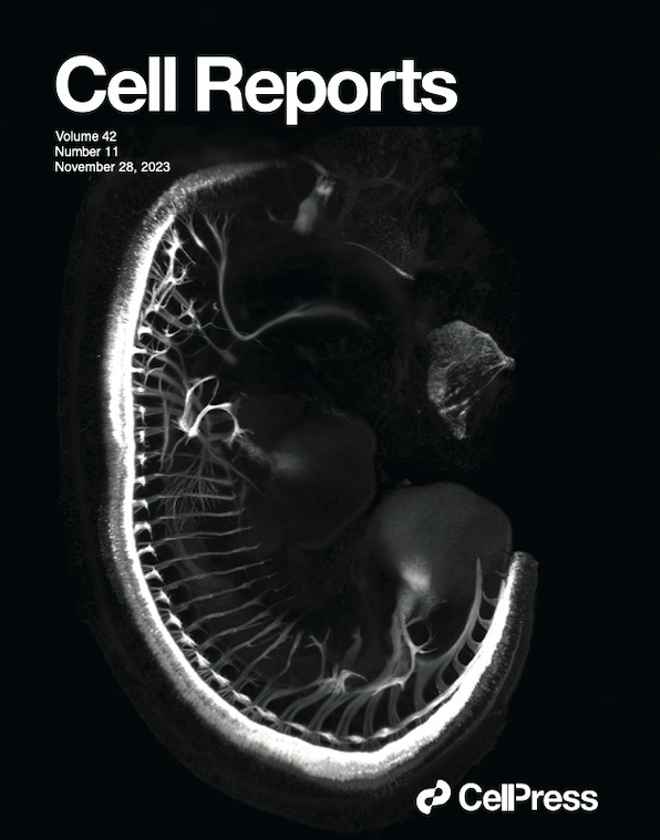
“Our collaboration with Milan and Jan’s teams was absolutely fundamental for performing the light-sheet microscopy and for all the image segmentation and quantifications used throughout the paper. The quality of the imaging data has been recognized by all three reviewers, one even praising it as "masterful". Indeed, Milan's imaging facility is the most excellent I have ever used. I am still amazed how much support, prompt help and encouragement we have received from his team,” says Alena. “Moreover, Cell Reports magazine selected one of our images of a mouse embryo as a cover photo for the November issue (see right).”
When asked if she plans to continue to collaborate with Milan’s facility, Alena’s answer was positive. She has already applied for a Czech BioImaging grant to support her next imaging project. “This time I plan to go in person,” says Alena enthusiastically.
Global BioImaging Advance the Conversation on Sustainable Biomedical Imaging Facilities
How can biomedical imaging facilities remain scientifically excellent while becoming more financially, environmentally, and operationally sustainable? These questions were at the heart of…
Moara Lemos: New insights from Global BioImaging’s Job Shadowing programme
Moara Lemos is a Brazilian researcher and imaging scientist. Her award winning research pushes the boundaries of structural biology, using advanced cryo-electron microscopy…
A Multiplexed Vision: Highlights from the Special Edition Virtual Pub on Spatial Transcriptomics
On June 12th, Euro-BioImaging hosted its latest Special Edition Virtual Pub, bringing together over 200 researchers and technology developers working at the cutting…