This review article by Lucien Hinderling and colleagues of the Euro-BioImaging Smart Microscopy Working Group, published in Methods in Microscopy in February 2026, provides a comprehensive overview of the rapidly evolving field of smart microscopy, in which image analysis and feedback control are integrated directly into the imaging process. The authors describe how adapting acquisition parameters such as magnification, illumination or region of interest for photomanipulation in real-time and based on detected biological events can increase throughput, improve reproducibility and enable automated experiments that would be impractical or impossible with predefined imaging workflows. The publication compiles representative use cases and technical strategies, highlighting common challenges and converging solutions.
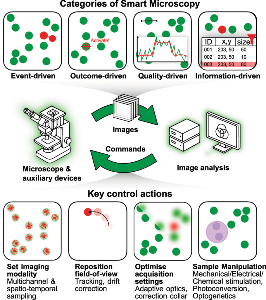
Categories and capabilities of smart microscopy systems integrating real-time image analysis and feedback control. Figure taken from Hinderling et al. (DOI: 10.1515/mim-2025-0029) under CC BY 4.
Workflows for developers
Lucien, a PostDoc at the University of Bern, tells how new technical developments in imaging and machine learning enable new ways to do science, citing an example from his own work:
“In the Pertz Lab, we use optogenetics to control cellular behavior. For example, you can illuminate a cell’s edge and watch it start migrating in a specific direction. But we had to select stimulation targets manually, which severely limited throughput. My PhD project was to build a smart microscope that uses computer vision to automate this targeting. I remember the first Monday after the microscope had run autonomously all weekend. We gathered around the screen and there was this massive dataset that would have taken us weeks to produce. But what excites me most is that smart microscopes are not just tools for automating existing workflows: to react to the sample in real time enables entirely new experimental paradigms, new ways to probe causal relationships or engineer artificial biological behaviors. Breakthroughs in machine learning have made such approaches possible; as we continue applying rapid advances in AI to microscopy, experiments will grow quickly in complexity.”
Workflows for facilities
But making smart microscopy more accessible does not just help an individual researcher to design complex workflows for a specifically challenging imaging application. Rafael Camacho, co-chair of the working group and one of the corresponding authors, has been developing workflows for a wide range of users at the Centre for Cellular Imaging at the University of Gothenburg for years. Fragmentation and a lack of interoperability between different commercial systems are key barriers to broader adoption.
“From a facility perspective, overcoming the current challenges in smart microscopy is essential because core facilities rely heavily on commercial instrumentation. We depend on robust, service-supported systems to guarantee reliability, quality, and long-term sustainability. At the same time, the era of data-driven research requires increasing flexibility, particularly at the software level, to implement adaptive and feedback-driven imaging workflows”, explains Rafael. “Facilities do not aim to rebuild microscope control software from scratch. Instead, we need well-defined communication layers that allow external software agents to interact with commercial systems. This would enable us to orchestrate experiments, integrate real-time image analysis, and implement intelligent feedback mechanisms while still operating within vendor-supported environments.”
Workflows for users
Aliaksandr Halavatyi, co-chair of the working group and second corresponding authors, not only provides access to instruments at the Advanced Light Microscopy Facility at EMBL, but also trains users in their manual and automated operation. He outlines some of existing challenges: “The implementations of smart microscopy workflows developed in recent years for commercial systems open the possibility to use this methodology by regular facility users who are non-experts in microscopy. However, making use of advanced microscopy workflows ultimately requires intensive user training and additional commitment of facility staff for configuring such experiments. Several standardised workflows can be identified, still each experiment might incorporate additional logic to make it most useful for the research project.”
Smart microscopy for all
The authors propose a roadmap centred on shared standards, modular software design and community-driven development. As Aliaksandr explains: “Powerful smart microscopy pipelines use different high-content imaging and photomanipulation modalities. Flexible combination of available acquisition and sample manipulation units and effective incorporation of available machine learning tools are essential for making smart microscopy workflows flexible and quickly adoptable to the needs of different projects. As existing tools for smart microscopy often rely on particular hardware and software combinations, re-using these workflows by different infrastructures or moving user projects from one microscope to another require expert knowledge and time commitment. Therefore, collaboration between academia and industry to develop interoperable modules and workflow tools for smart microscopy is critical to facilitate broader use of smart microscopy in the imaging facilities and beyond.”
The article is a call to action for researchers, developers, and manufacturers to work toward an open and connected ecosystem for smart microscopy. The Smart Microscopy Working Group provides a great platform for this collaboration. As Rafael adds: “By working together, we can ensure that commercial systems remain robust and maintainable, while also becoming interoperable and flexible enough to support next-generation smart microscopy applications. Our article represents an important step in building this shared vision across academia and industry.”
The SMWG brings together imaging scientists, software developers, and industry partners to coordinate efforts toward standardization and interoperability in smart microscopy. As one of the expert groups of Euro-BioImaging, the SMWG curates shared resources, documents existing implementations, and fosters dialogue between tool builders and users. Its activities create a unique enabling environment in which academic innovation and industrial development meet, accelerating the translation of advanced, adaptive imaging approaches into widely usable solutions for the life sciences community.
Major EU funding for user access & AI development, staff training, data stewardship & many more exciting new services!
Euro-BioImaging ERIC is deeply grateful to announce that the European Union has entrusted our infrastructure with funding to shape the future of imaging and…
Euro-BioImaging is looking for an Operations Support Assistant at the Euro-BioImaging Statutory Seat in Turku, Finland, to support the day-to-day financial and administrative operations…