
March 16, 2022
Welcome Linda Chaabane, new Med-Hub Section Director
As of March 1, 2022, Euro-BioImaging has a new Section Director at the Med-Hub, picking up where Professor Silvio Aime left off after 2…
March 16, 2022
As of March 1, 2022, Euro-BioImaging has a new Section Director at the Med-Hub, picking up where Professor Silvio Aime left off after 2…

March 16, 2022
Euro-BioImaging is organizing an online User Forum on April 5, 2022, from 14:00-17:00 CEST. This event will highlight the importance of…

March 16, 2022
Euro-BioImaging is organizing an online User Forum on April 5, 2022, from 14:00-17:00 CEST. This event will highlight the importance of…
March 16, 2022
Euro-BioImaging is organizing an online User Forum on April 5, 2022, from 14:00-17:00 CEST. This event will highlight the importance of…

March 15, 2022
Euro-BioImaging is organizing an online User Forum on April 5, 2022, from 14:00-17:00 CEST. This event will highlight the importance of…

March 15, 2022
Euro-BioImaging is organizing an online User Forum on April 5, 2022, from 14:00-17:00 CEST. This event will highlight the importance of…

March 14, 2022
Imaging living plants has always been challenging. Most microscopes place the sample horizontally – while plants grow vertically. But scientists at Czech Republic’s Institute…
March 10, 2022
On Friday, March 18that 13:00 CET, Silvia Pujals of the Institute for Advanced Chemistry of Catalonia (IQAC-CISC) delivers a lecture on “Correlative super resolution…
March 8, 2022
Euro-BioImaging is proud to participate in the Horizon Europe funded ISIDORe project, led by ERINHA, that brings together 154 project partners from 32 countries…
February 18, 2022
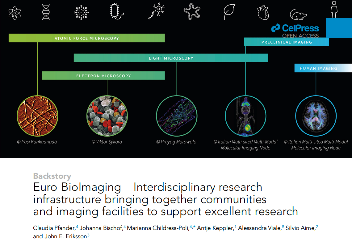
Euro-BioImaging has published an article “Euro-BioImaging – Interdisciplinary research infrastructure bringing together communities and imaging facilities to support excellent research” in open access…
February 17, 2022
What can we learn from the most comprehensive ELMI survey ever? In 2019, the European Light Microscopy Initiative community undertook one of the…
February 17, 2022
The Institut des Sciences Biologiques (INSB) of the French Centre National de la Recherche Scientifique (CNRS) has launched a call for projects to…