Need to look at the biomechanical properties of a sample? IPHYS Bioimaging Facility, part of Euro-BioImaging’s Advanced Light and Electron Microscopy Prague Node, offers Brillouin Microscopy in open access. This new service, which can be combined with spinning-disk fluorescent microscopy, is based on a commercial system the facility acquired through funding from the Czech-BioImaging modernization project. At a recent Brillouin Microscopy course at EMBL, we spoke to Šimon Vrana, an Imaging Specialist leading the biomechanics applications division of IPHYS BioImaging, to learn more about potential applications and how the service works from a user perspective.
Tell us who you are, where your facility is based, and what your role is.
My name is Šimon Vrana, and I’m an Imaging Specialist at BioImaging Facility, Institute of Physiology (IPHYS) of the Czech Academy of Sciences. Our facility is a part of both Czech-BioImaging and Euro-BioImaging. I am specialised in Atomic Force Microscopy, Brillouin and other biophysical methodologies. My role in the facility is to support our users with different imaging approaches to address scientific questions in this field of biomechanics.
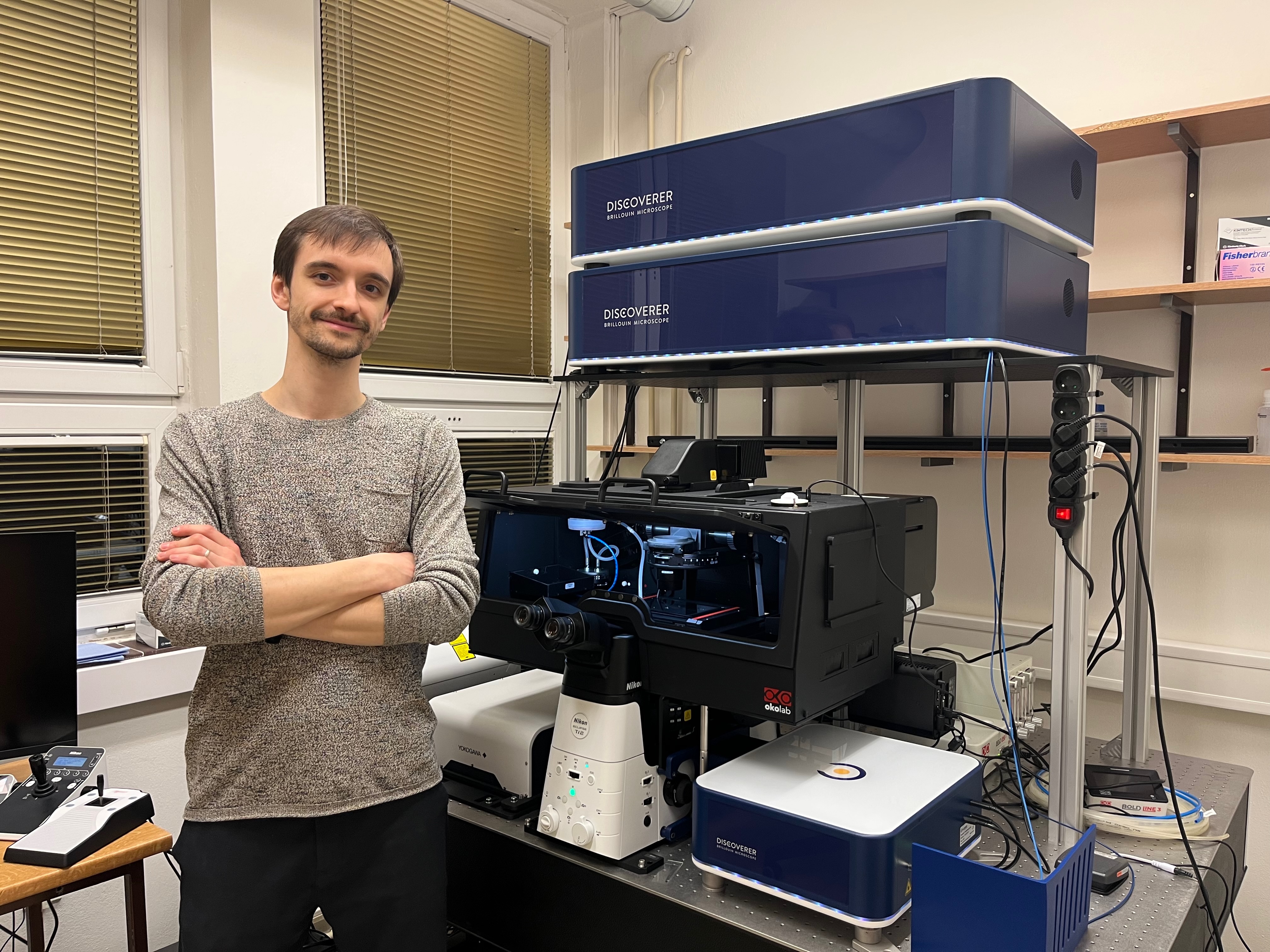
Šimon Vrana with the new Brillouin Microscope/Spinning Disk system at IPHYS Bioimaging Faciltiy, Czech Academy of Sciences, part of our Prague Node.
Your facility has recently invested in an off-the-shelf Brillouin Microscope. Please provide a short summary of what this microscope can do and list some applications:
Brillouin microscopy measures how a laser interacts with thermally driven acoustic waves in a sample, revealing information about the material’s internal mechanical characteristics and its resistance to deformation. Measuring properties such as longitudinal elastic modulus or viscoelastic behaviour in biological samples is crucial, since cells or tissues often differ in stiffness, and many pathological states are connected with changes of mechanical properties.
In the past, I worked primarily with cells and tissues, but the first users of our new Brillouin system are actually studying plants. We are currently measuring Physcomytrella moss samples provided by the Imaging Facility Institute of Experimental Botany ASCR. Specifically, one of the experiments involves comparing genetically modified Physcomitrella samples with their wild-type counterparts.
What kind of samples can you work with?
Brillouin microscopy was successfully used for measuring single cells, cell organoids, tissue samples, and plant samples. In general, we can examine any sample that can be viewed with confocal microscopy – the only condition is that the sample must be somewhat transparent and adherent. It is possible to measure non-adherent samples, but it is more difficult. Overall, sample preparation for Brillouin microscopy is usually not very demanding.
Why did you decide to invest in a commercial Brillouin system?
We chose the commercially available CellSense system for Brillouin Microscopy rather than a custom one because we want to offer Brillouin Microscopy as a service, and the CellSense system is very reliable and user-friendly. After a short training and some troubleshooting, users should be able to measure samples themselves. Commercial systems are still not widely available in Europe – we are currently the only Euro-BioImaging Node that is going to offer access to Brillouin modality. Even more, we can link Brillouin with spinning disk modality including Yokogawa super-resolution SoRa system. Here at the Brillouin course, we have been working with four systems, three of which are custom. Many custom systems are very good, but we don’t have enough resources and time to develop them. In addition, our user projects are very heterogenous, so the commercial system is the best option, and so far we’re satisfied with our decision.
What kind of projects do you hope to support using this technology? What scientific questions will you be able to address?
Our primary focus is on projects where biomechanics plays a central role in the biological question. We are particularly interested in supporting researchers who seek to understand how mechanical properties, such as stiffness, elasticity, and viscoelasticity, influence cellular behavior, tissue organization, and organismal development.
With Brillouin microscopy, we can address questions related to how mechanical properties change during development, differentiation, or disease progression. For example, we can study how stiffness varies within cell organoids, how mechanical gradients shape tissue morphogenesis, or how pathological states such as fibrosis, cancer, or neurodegeneration are associated with altered biomechanics. In plant biology, we can investigate how genetic modifications affect the mechanical integrity of cell walls or how environmental conditions influence tissue mechanics.
What other services do you provide in your facility that would be useful in combination with this type of microscopy?
Our setup is multimodal, combining Spinning Disk & Brillouin Imaging systems (learn more on our website). This integrated solution enables researchers to simultaneously capture high-resolution fluorescence dynamics of living cells and tissues, along with quantitative mechanical property mapping. It means that, for example, users can examine a cell sample stained with fluorescent dyes, identify a protein of interest, and then shift to Brillouin microscopy to measure local viscoelastic properties without dyes or mechanical probes. Data correlation is straightforward, and our facility provides full-service support for more complex data analysis.
Our facility offers a broad range of other modalities, including confocal microscopes, multiphoton-photon label-free modalities, FLIM, PLIM or light sheet, and we are proud to be able to correlate these modalities with compression, bi-axial or AFM experiments to help users in answering their research questions.
Why should scientists choose your facility for using this technology?
Scientists should choose our facility because we are one of a few Euro-BioImaging Nodes to offer Brillouin Microscopy in open access (Austrian BioImaging also offers Brillouin Microscopy in open access, learn more here as does France-BioImaging), and the only one to offer it with a commercial system that is linked to a confocal spinning disk system. We are very interested in integrating Brillouin microscopy into a multimodal workflow and our facility offersmany imaging modalities and fullsupport with the correlation.
In addition, we have a very comprehensive service offer, starting with consultancy services to help potential users to select the best imaging approach/modality to answer their scientific question. Once we have decided on an approach together with the user, we teach them to use the instrument and ensure they get the data they need.
We also provide support for image data analysis. Usually, commercial software, including one with the new Brillouin microscopy, offers a basic analysis. Beyond that, our facility assists users with third-party software packages such as Fiji, and we often implement personalized solutions in C++, C# or Python to analyze their data, if needed.
So, a great set-up, great service, and on top of that, Prague is a great city!
Many thanks for the interview!
About IPHYS BioImaging Facility
The IPHYS Bio-Imaging Facility is a member of the National Czech-Bioimaging infrastructure and a Prague node of Euro-Bioimaging with a 100 % open-access policy that makes advanced bioimaging techniques available to everyone!
Our team consists of biologists, engineers, and mathematicians who work toward accomplishing your scientific mission. The team provides help from designing the experiment, and data acquisition to data processing, analysis, and visualization.
Major EU funding for user access & AI development, staff training, data stewardship & many more exciting new services!
Euro-BioImaging ERIC is deeply grateful to announce that the European Union has entrusted our infrastructure with funding to shape the future of imaging and…
Euro-BioImaging is looking for an Operations Support Assistant at the Euro-BioImaging Statutory Seat in Turku, Finland, to support the day-to-day financial and administrative operations…