Peri implant connective tissue consistently forms an organised fiber arrangement around the implant neck during healing. Whithin this framework, X-ray phase-contrast (XPCT) imaging offers a suitable and raffinate tool to investigate the three-dimensional organization of connective tissue surrounding dental. abutments. This approach can help clarify the tissue's morphological architecture and shed light on how physical forces may influence this biological response during the wound-healing process.
Dr. Michele Furlani, from Polytechnic University of Marche (Italy), accessed the Phase Contrast Imaging (PCI) Flagship Node in Trieste (Italy) to study 30 surgical samples of connective tissue around dental implant abutments after three months of healing. The aim was to evaluate how implant geometry affects the healing process.
At the PCI Node, Dr. Furlani explored, on a larger and statistically significant sample size, the physical forces acting during the wound healing at the interface between connective tissue and dental implant abutment. He focused on both the macro- and micro-geometry of abutments to understand the microarchitecture of peri-implant soft tissues and the overall kinetics of the healing process. A set of structural parameters was quantified and compared using appropriate statistical tools.
Dr. Furlani demonstrated that clinical outcomes during peri-implant wound healing are influenced by the macro- and micro-geometry of the abutment, most likely determining special force settings at the interfaces between soft tissues and abutment. The quantitative analysis revealed a clear positive effect of the convergent geometry compared to the divergent one, with significantly greater amounts of connective tissue observed around both convergent groups. Moreover, abutments with micro-grooved surfaces outperformed machined ones in terms of collagen bundle intertwinement and density. An important implication of these findings is that collagen quantity and collagen fiber distribution can be considered independent quality factors in evaluating healing tissue.
Synchrotron 3D reconstructions of representative subvolumes (each with a fixed dimension of 540 × 270 × 270 μm3 and with the longest side of the prism as close as possible to the interface with the abutment) of the (a) DIV-MAC, (b) CONV-MAC, and (c) CONV-UTM groups: The decreased contrast between collagen fibers and interstitial spaces in the CONV-UTM group is an indication of an increased amount and interlacing of the collagen bundles. Figure has been adapted from Canullo et al, 2023: https://doi.org/10.1111/clr.14118. Used under a Creative Commons CC-BY 4.0 licence.
These results also invite further reflection on the biomechanics underlying such morphological responses: the structural forces exerted during healing strongly depend on the surrounding geometrical conditions, which in turn influence both the directionality and the morphological complexity of the forming connective tissue. Awareness of these physical responses can therefore provide valuable insights into tissue stability.
Read more:
Case Report of a Dental Implant with Conometric Abutment–Prosthetic Cap Connection: Advanced High-Resolution Imaging and Peri-Implant Connective Tissue Performance https://doi.org/10.3390/clinpract14020043
Influence of abutment macro- and micro-geometry on morphologic and morphometric features of peri-implant connective tissuehttps://doi.org/10.1111/clr.14118
About Euro-BioImaging’s Phase Contrast Imaging (PCI) Flagship Node
The Italian Phase Contrast Imaging Flagship Node is based on the SYRMEP beamline of the Elettra Synchrotron light source (Trieste). The main characteristics of Synchrotron Radiation, namely monochromaticity, high intensity and spatial coherence, allow the effective application of phase contrast techniques. Differently from conventional radiology, where the image formation relies on the absorption properties of the sample, these approaches are sensitive to the phase shifts produced by the sample on the incoming X-rays. Phase contrast is particularly effective for imaging of soft biological tissues, where the conventional technique has strong limitations due to the poor intrinsic X-ray absorption. The beamline provides two stations working with monochromatic or white/pink X-ray beam for planar and Computed micro-Tomography (microCT) imaging. A CT system based on a micro-focus X-ray source, named TomoLab, is available, if required in the proposal, as auxiliary facility. The Node offers full packages including image acquisition, reconstruction and data reduction tools.
More news from Euro-BioImaging
March 24, 2026
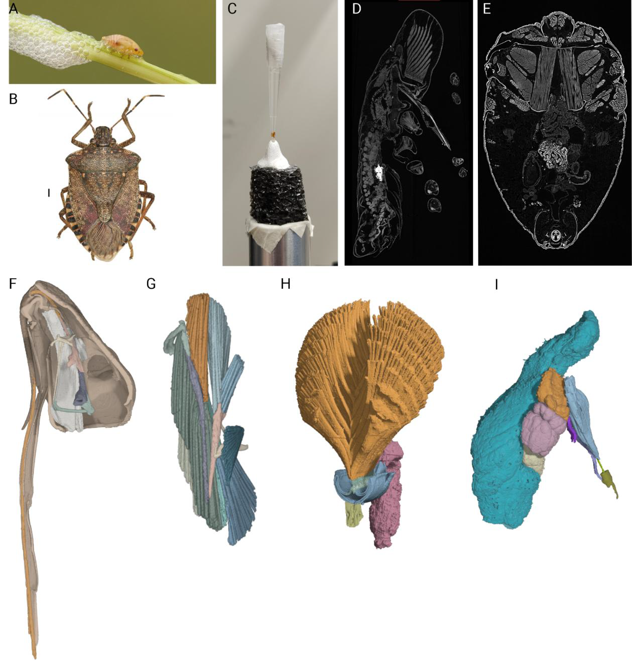
Use of synchrotron X-ray Phase-contrast MicroCT to investigate the reproductive system and the structures involved in foam-production in the spittlebug Philaenus spumarius
Use of synchrotron X-ray Phase-contrast MicroCT to investigate the reproductive system and the structures involved in foam-production in the spittlebug Philaenus spumarius The Olive…
France BioImaging Annual Meeting 2026 highlights leadership, collaboration and scientific excellence
The 2026 Annual Meeting of France BioImaging, held on 12–13 March at the University of Rouen Normandie, brought together Node leaders, facility staff, industry…
EVOLVE Distributed In-Person Training Course: Intro to BioImaging Analysis with Python for Life Scientists
After a very successful first edition, Euro-BioImaging is proud to announce the second edition of the distributed training course for “Intro to BioImage Analysis…