
March 30, 2026
Euro-BioImaging at the EOSC ESFRI meeting in Milan
Euro-BioImaging was delighted to attend the ESFRI/EOSC Policy Workshop on “EOSC and Research Infrastructures: Opportunities and Strategies” in Milan from March 15-16, represented by…

Contrast agents (CA) for magnetic resonance imaging (MRI) are widely used in clinical practice to improve diagnostic accuracy. Despite the success of gadolinium (III)-chelate-based CAs due to their excellent relaxation properties, some safety concerns have been raised.
Dr. Lucio Melone (Politecnico di Milano, Italy) developed a set of cyclodextrins water soluble contrast agents functionalized with different kinds of nitroxide radicals to be tested both in vitro and in vivo at the Finnish Biomedical Imaging Node.
In vivo MRI experiments at 9.4 T in a glioma rat model showed a good relaxivity and an improved retention of the contrast for at least 60 minutes, confirming an improved stability of cyclodextrins nitroxide radicals also in vivo.
Read more here:
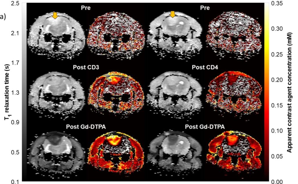
The Finnish Biomedical Imaging Node (FiBI) is a multi-sited, multimodal Node covering biomedical imaging from mouse to man. The spearhead imaging technologies of the FiBI Node include 1) preclinical and human PET imaging and PET tracer development, 2) preclinical high-field MRI, 3) magnetoencephalography (MEG), and 4) optical intravital imaging, coupled with a broad repository of imaging tracers and probes, numerous animal models from mice to pigs, and diverse stimulation systems for both animals and humans. The key expertise and main research applications focus on major challenges especially in cardiovascular and metabolic diseases, neuroscience, and cancer. With wide coverage of imaging modalities and expertise, the FiBI Node provides exceptional opportunities not only for basic research but also for translational research from small animals to larger animals to humans and to the clinic within a single Node.
March 30, 2026
Euro-BioImaging was delighted to attend the ESFRI/EOSC Policy Workshop on “EOSC and Research Infrastructures: Opportunities and Strategies” in Milan from March 15-16, represented by…
March 30, 2026
Euro-BioImaging was delighted to attend the Public Awareness & Engagement of Research Infrastructures (PAERI) conference, represented by External Communications Officer, Marianna Childress-Poli. This year’s…

March 26, 2026
The Madrid Advanced Microscopy Center (MAdMiC) is the first Euro-BioImaging Node in Madrid (Spain). It is formed through the collaboration and close work of…