Metabolic dysfunction-associated steatotic liver disease (MASLD), formerly known as non-alcoholic fatty liver disease (NAFLD), is the most prevalent liver disease globally, yet no therapies are approved.
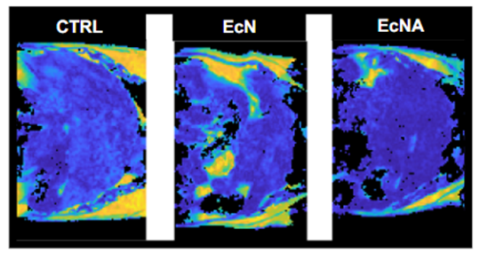
Dr Johnson Lok, from the University of Eastern Finland, was interested in understanding the effects of Escherichia coli Nissle 1917 strains, engineered to express human hormones, as potential hormonal and microbial therapy for the disease. To do so, he applied for access at the Finnish Biomedical Imaging Node, Kuopio Biomedical Imaging Unit, to use Magnetic Resonance Imaging (MRI) to measure the extent of liver fat accumulation in a mouse model of MASLD. This study demonstrated that the treatment with the engineered E. coli Nissle strain, along with dietary changes, reduced body weight, liver steatosis, and plasma cholesterol levels, supporting the potential for using engineered microbial therapeutics in combination with dietary changes for managing the disease.
Representative MRI images of livers. A lighter colour indicated a higher MRI fat index which means a higher liver fat content. Figure adapted from Iannone et al. 2023. Used under a Creative Commons CC-BY 4.0 licence.
Read more:
Changes in liver metabolic pathways demonstrate efficacy of the combined dietary and microbial therapeutic intervention in MASLD mouse model - https://doi.org/10.1016/j.molmet.2023.101823
Advanced Microbiome Therapeutics Accelerate MASLD Recovery by Restoring Intestinal Microbiota Equilibrium and the Gut-Liver Axis in a Mouse Model - https://doi.org/10.1021/acs.jafc.5c01674
About the Finnish Biomedical Imaging (FiBI) Node
The Finnish Biomedical Imaging Node (FiBI) is a multi-sited, multimodal Node covering biomedical imaging from mouse to man. The spearhead imaging technologies of the FiBI Node include 1) preclinical and human PET imaging and PET tracer development, 2) preclinical high-field MRI, 3) magnetoencephalography (MEG), and 4) optical intravital imaging, coupled with a broad repository of imaging tracers and probes, numerous animal models from mice to pigs, and diverse stimulation systems for both animals and humans. The key expertise and main research applications focus on major challenges especially in cardiovascular and metabolic diseases, neuroscience, and cancer. With wide coverage of imaging modalities and expertise, the FiBI Node provides exceptional opportunities not only for basic research but also for translational research from small animals to larger animals to humans and to the clinic within a single Node.
More news from Euro-BioImaging
April 9, 2026
Major EU funding for user access & AI development, staff training, data stewardship & many more exciting new services!
Euro-BioImaging ERIC is deeply grateful to announce that the European Union has entrusted our infrastructure with funding to shape the future of imaging and…
Euro-BioImaging is looking for an Operations Support Assistant at the Euro-BioImaging Statutory Seat in Turku, Finland, to support the day-to-day financial and administrative operations…