
April 7, 2026
Euro-Bioimaging attends the EMIM 2026 Conference
We were delighted to take part in the 21st European Molecular Imaging Meeting (EMIM), held in Ljubljana, Slovenia, from March 24–27, 2026. The conference…

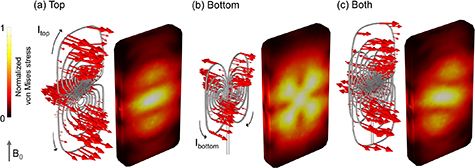
Transcranial magnetic stimulation (TMS) is a non-invasive method for stimulating the cerebral cortex. Concurrent functional magnetic resonance imaging (MRI) can show changes in TMS-induced activity in the whole brain, with the potential to inform brain function research and to guide the development of TMS therapy. However, the interaction of the strong current pulses in the TMS coil with the static main magnetic field of the MRI scanner produces high Lorentz forces, which may damage the coil enclosure and compromise the patient's safety.
Maria Koponen, during her master’s thesis, studied the mechanical stresses of rat multi-locus TMS (mTMS) transducers inside a high-field MRI scanner (9.4T), using computational modeling and wanted to test some material options in a real fMRI-environment. In collaboration with the Finnish Biomedical Imaging Node, Kuopio Biomedical Imaging Unit, she studied the time-dependent mechanical behavior and durability of two selected mTMS coil arrays. She found that the current pulses produce shock waves and time-dependent stress distribution in the coil plates, whose intensity and location depend on the current waveform, the coil combination, and the transducer orientation relative to the MRI magnetic field. This study allowed her to define the most durable material, out of the six options studied, and provide novel insights for more durable TMS coil designs , which are essential for developing mechanically stable and safe mTMS-MRI transducers.
About the Finnish Biomedical Imaging (FiBI) Node
The Finnish Biomedical Imaging Node (FiBI) is a multi-sited, multimodal Node covering biomedical imaging from mouse to man. The spearhead imaging technologies of the FiBI Node include 1) preclinical and human PET imaging and PET tracer development, 2) preclinical high-field MRI, 3) magnetoencephalography (MEG), and 4) optical intravital imaging, coupled with a broad repository of imaging tracers and probes, numerous animal models from mice to pigs, and diverse stimulation systems for both animals and humans. The key expertise and main research applications focus on major challenges especially in cardiovascular and metabolic diseases, neuroscience, and cancer. With wide coverage of imaging modalities and expertise, the FiBI Node provides exceptional opportunities not only for basic research but also for translational research from small animals to larger animals to humans and to the clinic within a single Node.
Read more: Modeling the stress and forces on multi-channel TMS coil arrays in high-field MRI scanners - https://doi.org/10.1088/1361-6560/ad6b72
April 7, 2026
We were delighted to take part in the 21st European Molecular Imaging Meeting (EMIM), held in Ljubljana, Slovenia, from March 24–27, 2026. The conference…
April 7, 2026
Following the continued success of the Scientific Ambassadors programme, Euro-BioImaging is delighted to welcome its 2026 cohort. Building on the strong foundation laid by…

March 30, 2026
Euro-BioImaging was delighted to attend the ESFRI/EOSC Policy Workshop on “EOSC and Research Infrastructures: Opportunities and Strategies” in Milan from March 15-16, represented by…