BioImage data analysis is a fast-paced field, at the intersection of computer science and other scientific disciplines. BioImage data analysts support user projects from areas of research as diverse as mechanobiology, microbiology, histocytochemistry, plant science and material science. In a field that defies disciplines, how do bioimage data analysts stay cutting-edge? How do they hone their skills and strengthen their networks? At training courses like the EMBO course on Advanced methods in BioImage Analysis, that took place at EMBL Heidelberg from September 14-19, 2025, with attendees from multiple Euro-BioImaging Nodes and image analysts from around the world.
ORGANISERS:
The course was organised by Ignacio Arganda-Carreras, University of the Basque Country (UPV/EHU, Bilbao Node), Anna Klemm, Scilifelab and Uppsala University (Swedish Node), Perrine Paul-Gilloteaux, University of Nantes (France-Bioimaging) and Christian Tischer, EMBL Heidelberg together with Julia Patricia Nohle and Irena Provaznikova, from EMBL-Events, EMBL Heidelberg.
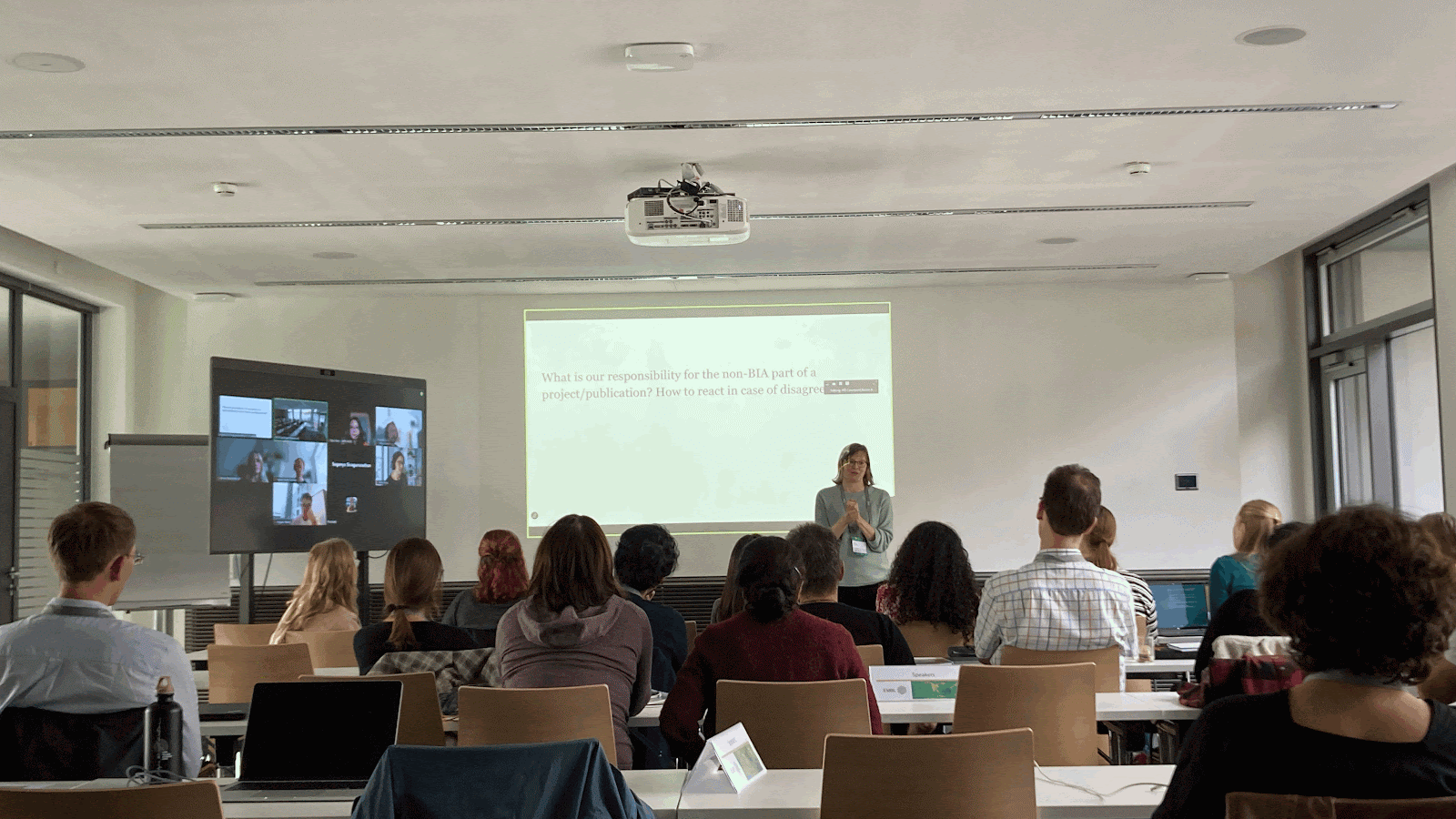
“The privilege of our job is to never be bored,” said Jean-Yves Tinevez, Institut Pasteur and France-BioImaging, referring to the diversity of projects that Core Facility Image Analysts work on. His comment reflected the general enthusiasm of the room, during a discussion session at the EMBO Course. The discussion was moderated by Anna Klemm, SciLifeLab & Uppsala University, part Euro-BioImaging’s Swedish National Microscopy Infrastructure (NMI), co-organiser of the course. She gathered insights from image analysts hailing from several Euro-BioImaging Nodes and other research institutions across Europe, but also Singapore, Ecuador, USA, and Israel, some of whom attended online.
Anna Klemm, SciLifeLab and Uppsala University, Swedish Node, leads the discussion on the role of image analysts in core facilities.
Training the experts
“A majority of attendees are core facility staff, to ensure maximum impact,” explained Perrine Paul-Gilloteaux, University of Nantes, France, part of Euro-BioImaging’s France-BioImaging Node and co-organiser of the course. “The people attending this course will go home and share what they learned with many others at their home institute. The best way to disseminate knowledge about image analysis tools is to train the analysts themselves. They will ensure that the maximum number of researchers benefit from the expertise.”
The intensive week-long training course provided multiple opportunities for hands on learning, but also discussion. Experts discussed the dilemma of working with commercial image analysis software or open source, and shared their thoughts on the responsibility of image analysts for data quality or scientific interpretation.
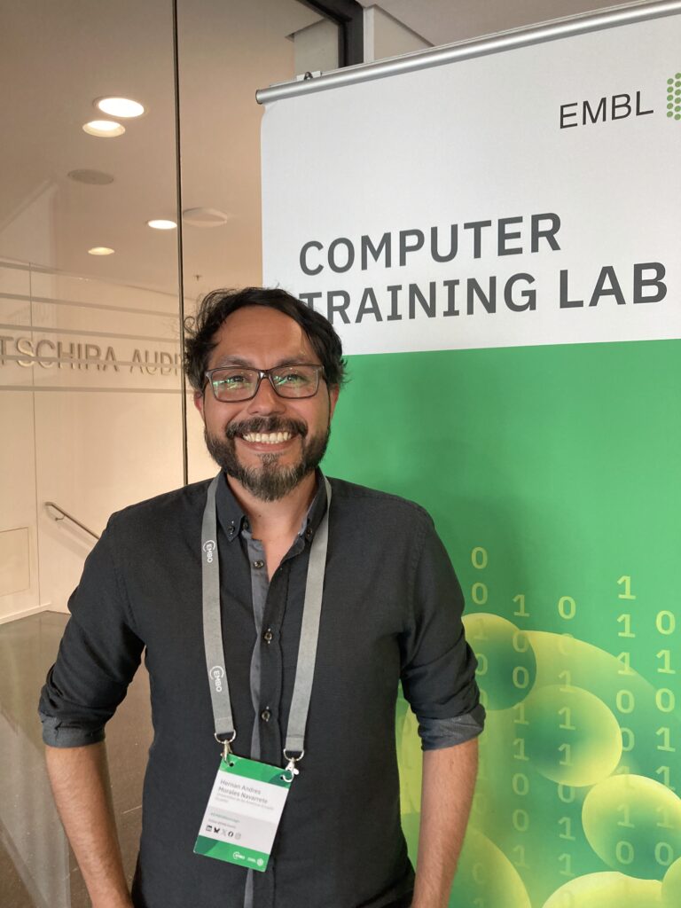
“I am really happy to have had the opportunity to attend this course, thanks to the I4A funding. I not only learned about cutting-edge tools and pipelines but also built a strong network of collaborators for future projects."
-- Hernán Andrés Morales-Navarrete, Universidad de las Américas
Sharing best practices
Image Analysis Core Facilities are super important because users (researchers) come to the image analysts to get advice on image analysis even before they start imaging. “Having the people who will do the analysis give advice at the beginning of the project is extremely important for the end result,” said Nicolas Peredo, image analyst at VIB/KU Leuven, part of Flanders BioImaging. “We really encourage our users to talk to us first - it’s a best practice. At first there was a bit of a learning curve. Now the users themselves come to us before their project starts, and tell other researchers to do so, too, because the results are so much better.”
Learning new software & working with AI
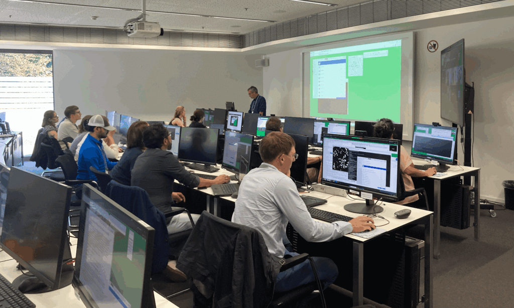
Sessions covered topics ranging from workflows and data management to the use of Artificial Intelligence in Image Analysis, spatial statistics, spatial omics, force modelling and more. A number of different software tools were explored. The format was following the concept of the flipped classroom with on-site sessions dedicated to practicals.
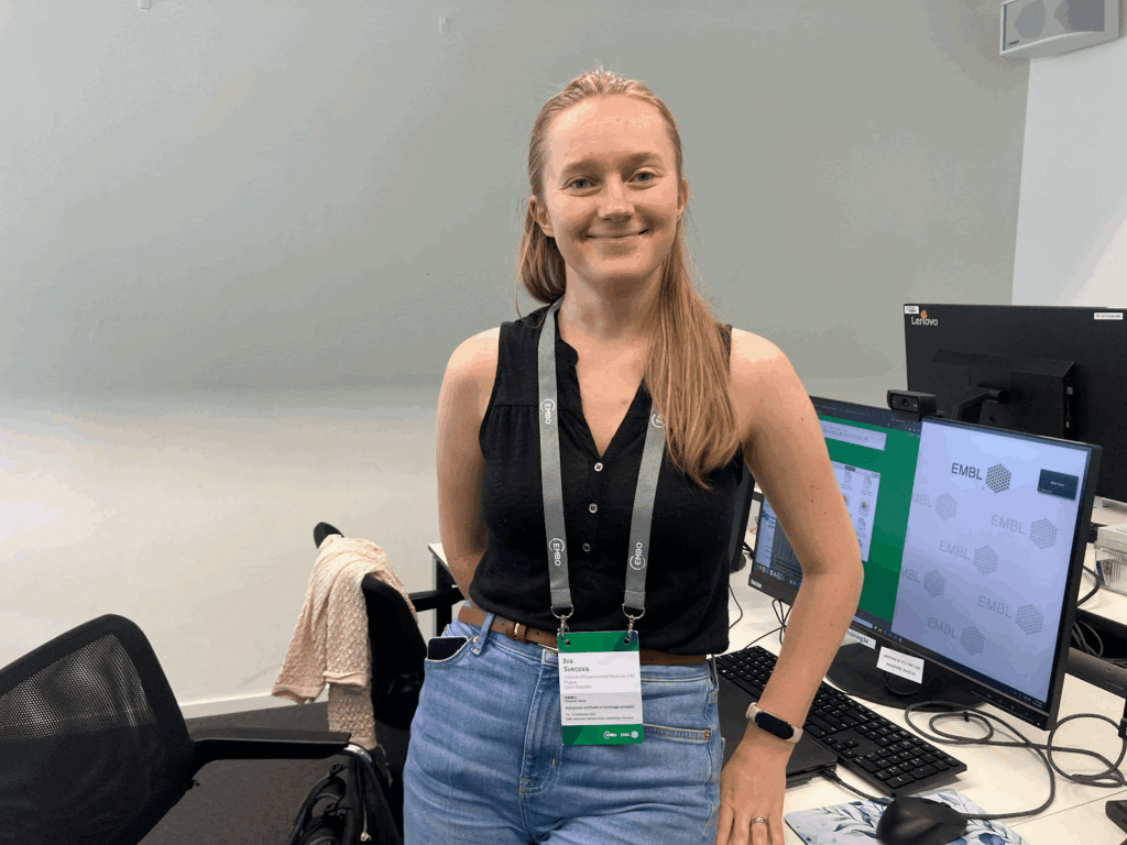
“The course was super beneficial in many aspects. First, we learned a lot about various bioimage analysis tools, some of which (such as Nextflow or BiaPy) were already on my curiosity list. There was also plenty of time to discuss with others and share our experience, which increased the learning opportunities and gave me a lot of thoughts on how to improve services in our facility.”
-- Iva Svecova, IEM-CAS
An international group
Getting into the course was quite competitive, and the students came from geographically diverse parts of the globe. Hernán Andrés Morales-Navarrete attended the Advanced Methods in Bioimage Analysis course, representing the Universidad de Las Américas in Ecuador. Hernán is a physicist developing machine learning applications and physics-based computational models for biological systems – focused on embryonic development and liver tissue analysis.
“What I enjoyed most about this course was the opportunity to learn hands-on about state-of-the-art bioimage analysis methods, directly from the top experts in the field. I especially appreciated the sessions on open-source tools and the FAIR data principles, which strongly align with my commitment to open science and reproducible research. The interactive format of the course, with practical sessions and direct discussions with instructors and attendees, was incredibly valuable.”
His trip was funded by Global BioImaging’s Imaging 4 All initiative, which supports researchers from low- and middle-income countries (LMICs) in accessing advanced imaging technologies.
Hernán Andrés Morales-Navarrete attended the Advanced Methods in Bioimage Analysis course, representing the Universidad de Las Américas in Ecuador, with funding from Global BioImaging's Imaging 4 All initiative.
“I am really happy to have had the opportunity to attend this course, thanks to the I4A funding. I not only learned about cutting-edge tools and pipelines but also built a strong network of collaborators for future projects. I’m also planning to organise a Bioimage Analysis course in Latin America in 2026, and the way this course is run has been very inspirational, giving me concrete ideas of what I can implement back home to strengthen the local community.’
Iva Svecova, from IEM CAS, part of Euro-BioImaging’s Advanced Light & Electron Microscopy Prague Node, also attended the course, thanks to a grant from the EVOLVE project, that supports Euro-BioImaging Node staff wanting to attend training courses at imaging facilities anywhere in the world. “I’m really happy to have received this funding because without it, I wouldn’t have been able to attend,” explains Iva. “The course was super beneficial in many aspects. First, we learned a lot about various bioimage analysis tools, some of which (such as Nextflow or BiaPy) were already on my curiosity list. There was also plenty of time to discuss with others and share our experience, which increased the learning opportunities and gave me a lot of thoughts on how to improve services in our facility.”
Iva Svecova, from IEM CAS, part of Euro-BioImaging’s Prague Node, attended the course with EVOLVE funding.
“Moreover, I really enjoyed the social aspect of the course, as everybody was very enthusiastic and had ideas to share. Many of the organisers and speakers are active in the bioimage analysis community, and after knowing their names from webinars and publications, it was amazing to finally get to know them in person.“
This course provided thus important opportunities for image analysts to hone their skills and build their networks, building talent, sharing the best tools, and creating an interconnected web of image analysts to face the challenges of the fast-evolving image data analysis field.
Studious hands on session about Statistical spatial analysis and modeling with BIP, led by Philippe Andrey, INRAE Versailles, France.
More news from Euro-BioImaging
April 7, 2026
canSERV User Meeting highlights impact and future perspectives for cancer research in Europe
The canSERV Annual Meeting, held in Brussels from 25–27 March 2026, brought together researchers, service providers, Research Infrastructures, policymakers, and patient representatives to reflect…
We were delighted to take part in the 21st European Molecular Imaging Meeting (EMIM), held in Ljubljana, Slovenia, from March 24–27, 2026. The conference…
Euro-BioImaging Welcomes its 2026 Scientific Ambassadors Cohort
Following the continued success of the Scientific Ambassadors programme, Euro-BioImaging is delighted to welcome its 2026 cohort. Building on the strong foundation laid by…