
March 30, 2026
Euro-BioImaging at the EOSC ESFRI meeting in Milan
Euro-BioImaging was delighted to attend the ESFRI/EOSC Policy Workshop on “EOSC and Research Infrastructures: Opportunities and Strategies” in Milan from March 15-16, represented by…

Breast cancer remains one of the most common cancers worldwide, and HER2-positive tumours — characterised by overexpression of the HER2 receptor — are known for their aggressive behaviour and complex treatment needs. Developing therapies that are both effective and highly targeted is therefore a major research priority. Through the EU-funded canSERV project (GA# 101058620), researchers across Europe are gaining access to cutting-edge Research Infrastructure services to accelerate cancer research and innovation. Euro-BioImaging is a core partner in canSERV, providing open access to advanced biological and biomedical imaging facilities and expertise to support cancer researchers from across Europe. Within this framework, a multidisciplinary research team from Slovakia explored a novel approach to HER2-positive breast cancer treatment by combining protein engineering with advanced imaging — supported by the Cellular Imaging Hungary Node of Euro-BioImaging.
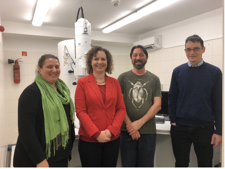
Veronika Huntošová and her team from the Center for Interdisciplinary Biosciences at Technology and Innovation Park of the Pavol Jozef Šafárik Univeristy in Košice are working to develop new breast cancer therapies. Her canSERV project focused specifically on the early development of a highly selective photodynamic therapy approach. Photodynamic therapy relies on light-activated molecules — photosensitisers — that can trigger cell damage when illuminated, but its clinical use depends critically on delivering these molecules specifically to cancer cells.
To address this, the researchers designed a genetically encoded photosensitising protein that can be guided directly to HER2 receptors on breast cancer cells. By combining a light-responsive protein domain with a targeting molecule that recognises HER2, the team created a system intended to deliver the photosensitiser selectively inside tumour cells, minimising off-target effects. This work represents an important preparatory step towards more precise and personalised treatment strategies for HER2-positive breast cancer, where targeted delivery is key.
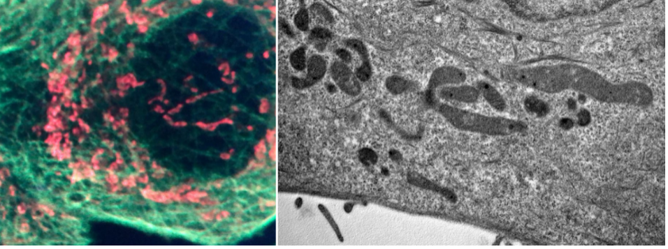
Advanced imaging played a central role in understanding how the engineered protein behaves once it encounters cancer cells. At the Cellular Imaging Hungary Node site at the University of Debrecen, the team accessed complementary light and electron microscopy services to visualise the interaction between the targeting protein and breast cancer cells at multiple levels.
Live-cell and high-resolution fluorescence microscopy allowed researchers to observe how the protein recognises HER2 receptors, enters cells, and affects key cellular structures. Electron microscopy then provided ultrastructural insight, revealing changes in mitochondrial organisation — an early indicator of how cells respond to the treatment concept.
Together, these imaging approaches helped the team build a coherent picture of how the targeted photosensitiser primes cancer cells ahead of future photodynamic treatment, both in conventional cell cultures and in more advanced 3D cancer models planned for follow-up studies.
While this study focused on cellular-level validation rather than clinical application, its results form a strong foundation for the next stages of research. The findings will inform ongoing and future projects exploring targeted photodynamic therapy in breast cancer organoids and other advanced models, as well as a planned high-impact scientific publication.
Equally important, the project strengthened international collaboration and skills development. Multiple visits to the Cellular Imaging Hungary Node of Euro-BioImaging enabled hands-on training for Veronika and her students, close interaction with facility experts, and long-term scientific exchange — illustrating how shared European research infrastructures support both scientific progress and researcher development.
“Access to Euro-BioImaging through canSERV allowed us to combine advanced imaging techniques in ways that would not have been possible within a single laboratory. This was essential for understanding how our targeted therapy concept behaves inside cancer cells and for shaping the next phase of our research.”
— [Veronika Huntošová, principal investigator, Center for Interdisciplinary Biosciences at Technology and Innovation Park of the Pavol Jozef Šafárik Univeristy in Košice]
March 30, 2026
Euro-BioImaging was delighted to attend the ESFRI/EOSC Policy Workshop on “EOSC and Research Infrastructures: Opportunities and Strategies” in Milan from March 15-16, represented by…
March 30, 2026
Euro-BioImaging was delighted to attend the Public Awareness & Engagement of Research Infrastructures (PAERI) conference, represented by External Communications Officer, Marianna Childress-Poli. This year’s…

March 26, 2026
The Madrid Advanced Microscopy Center (MAdMiC) is the first Euro-BioImaging Node in Madrid (Spain). It is formed through the collaboration and close work of…