
July 9, 2026
Euro-BioImaging Visits the NORMOLIM Node at the 2026 180°N Conference
In April 2026, the Med-Hub Head of Operations Alessandra Viale attended the 2026 180°N Conference, held at the beautiful Nye Hjorten Teater…

One of Euro-BioImaging’s key missions is to facilitate excellent science by providing open access to imaging technologies and expertise. Users come from many backgrounds – and career stages – to work with core facility experts. But what about the experts themselves? What if they want to test a new technique or develop a new approach? One important aspect of Euro-BioImaging is bringing our Nodes together, so experts can learn from each other, try out new techniques – and expand their portfolio.
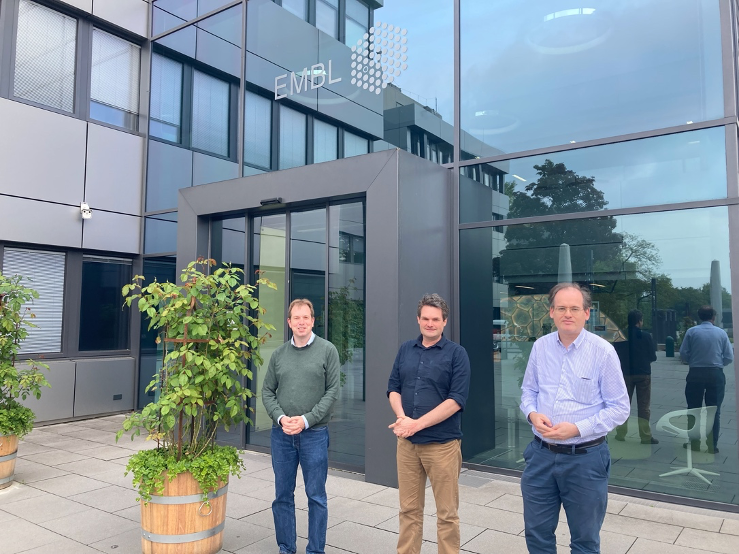
Dorus Gadella and Mark Hink work at the University of Amsterdam, and represent our Van Leeuwenhoek Center for Advanced Microscopy (LCAM) - Functional Imaging Flagship Node. They are experts in functional imaging and in probe development to advance quantitative life imaging of cellular processes at highest resolution. They recently developed a range of new red-light fluorescent protein probes for Stimulated emission depletion microscopy (STED) – a super-resolution technique that is currently not available at their facility. So, they applied for access at Euro-BioImaging’s EMBL Node - where different types of STED system are available. Their application was accepted – and in a highly competitive process, was also selected to receive funding from the Euro-BioImaging User Access fund.
New fluorescent probes for super resolution microscopy
“We have been developing new fluorescent protein probes with high fluorescence lifetimes for live cell imaging at super-resolution. We started with the best red fluorescent proteins then introduced mutations, to make them more photostable to enable super resolution,” explains Dorus Gadella, Professor in Molecular Cytology and Director of the LCAM.
“STED exposes cells to a lot of light and can kill them or impact their behavior – you therefore need probes that are super bright and resilient. We wanted to bring our new probes to the EMBL Node because it offers Tau STED, normal STED and RESOLFT. Having access to the Node allows us to test the probes performance on all these different systems” says Mark Hink, Assistant Professor and Manager of the van Leeuwenhoek Center for Advanced Microscopy. “The Euro-BioImaging User Access Fund was a catalyst that coincided with the development of our probes and gave us incentive to write this proposal.”
“Furthermore, we are considering investing in a STED system in Amsterdam,” says Dorus Gadella. “But we really wanted to test it first – to get a feeling for how it works with biologically relevant samples: live cells with fluorescent protein-labeled structures. Just to have the instrument – or to test it with the manufacturer – is not enough. You really need a lot of expertise. With this kind of instrument, it’s not just about pushing the buttons. You need to understand how the physics works and do a lot of finetuning of the settings. The exchanges we’ve had while sitting at the microscope with Marko, just being able to discuss with an expert spontaneously and informally, is unique.”
Working together – for the best scientific results
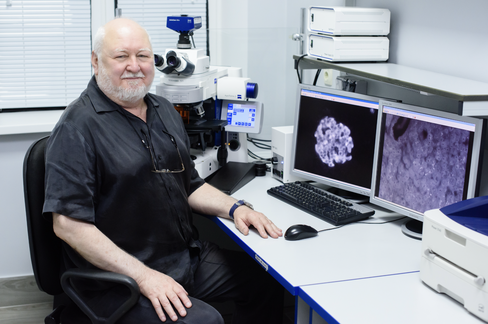
Marko Lampe is a Scientific Officer at EMBL’s ALMF – and an expert in STED microscopy. To prepare for Dorus and Mark’s visit, he transfected some cells so they would express the new fluorescent proteins fused to different cytoskeletal proteins that would be examined in the project, making the project more efficient and allowing experiments to start as soon as the visitors from Amsterdam arrived. “We enjoy having external visitors,” says Marko. “We reserve special time for them, and often really become immersed in their projects. But if you only have a week you have to be very efficient. Testing new probes is challenging but fun - it can take half a day just to optimize the settings.”
During their visit, the team from Amsterdam gathered a lot of STED data on their different probes to characterize their properties in STED applications.
Some riddles to unravel
“We are happy with our results – I now have a good feeling about what these different STED techniques can do. Our next step is to analyze all of the data and think deeply about our observations before making some further adapted probes. Then we will come back for another visit in the fall with more and hopefully even better probes,” explains Dorus.
But of course, no testing of new probes would be complete without some surprises. When they tested one of the new probes, they observed a strange change to the fluorescence lifetime only under STED illumination, which was an unexpected observation and will require further testing to understand the underlying photophysical processes.
The advantages of multiple instruments
“That’s where the Tau STED was very important,” says Marko. “With Tau STED, we get fluorescence lifetime information. On regular STED, without lifetime contrast, we would have just considered one fluorescent protein better than another, but had no clue about the mechanism, the Tau system showed that something was happening with the fluorescent lifetime of the probe.”
So, which protein is better? The puzzle remains for the next visit.
In the meantime, there are loads of data to analyze, and a protocol to develop. But the outset for the new probes is very promising. Which goes to show that bringing experts with different backgrounds together can lead to very exciting science - and advances in imaging applications. In this particular case, it could also result in expanding the technology portfolio of the LCAM Node to include STED. Mark and Dorus will return to the EMBL Node for a second visit to further evaluate their improved STED probes.
About the EMBL Node
The EMBL Node is made up of 4 facilities, the Advanced Light Microscopy Facility (ALMF) and the Electron Microscopy Core Facility (EMCF) at EMBL Heidelberg, the Mesoscopic Imaging Facility at EMBL Barcelona, and newly joined in May 2022, the EMBL Imaging Centre (EMBL IC). The cross-comparisons between different STED approaches that were the goal of Mark and Dorus’ visit, were made possible by using the instrumentation available at both the ALMF and the EMBL IC. The EMBL Imaging Centre, as the newest part of the EMBL Node, is dedicated to rapidly making the most advanced light and electron microscopy technologies available as a synergistic service portfolio to the international user community from both academia and industry to enable new ground-breaking research that crosses the scales of biology.
July 9, 2026
In April 2026, the Med-Hub Head of Operations Alessandra Viale attended the 2026 180°N Conference, held at the beautiful Nye Hjorten Teater…
July 7, 2026
Armed conflicts generate long-lasting environmental contamination that extends well beyond the duration of military operations. The release of heavy metals such as Arsenic,…
July 6, 2026
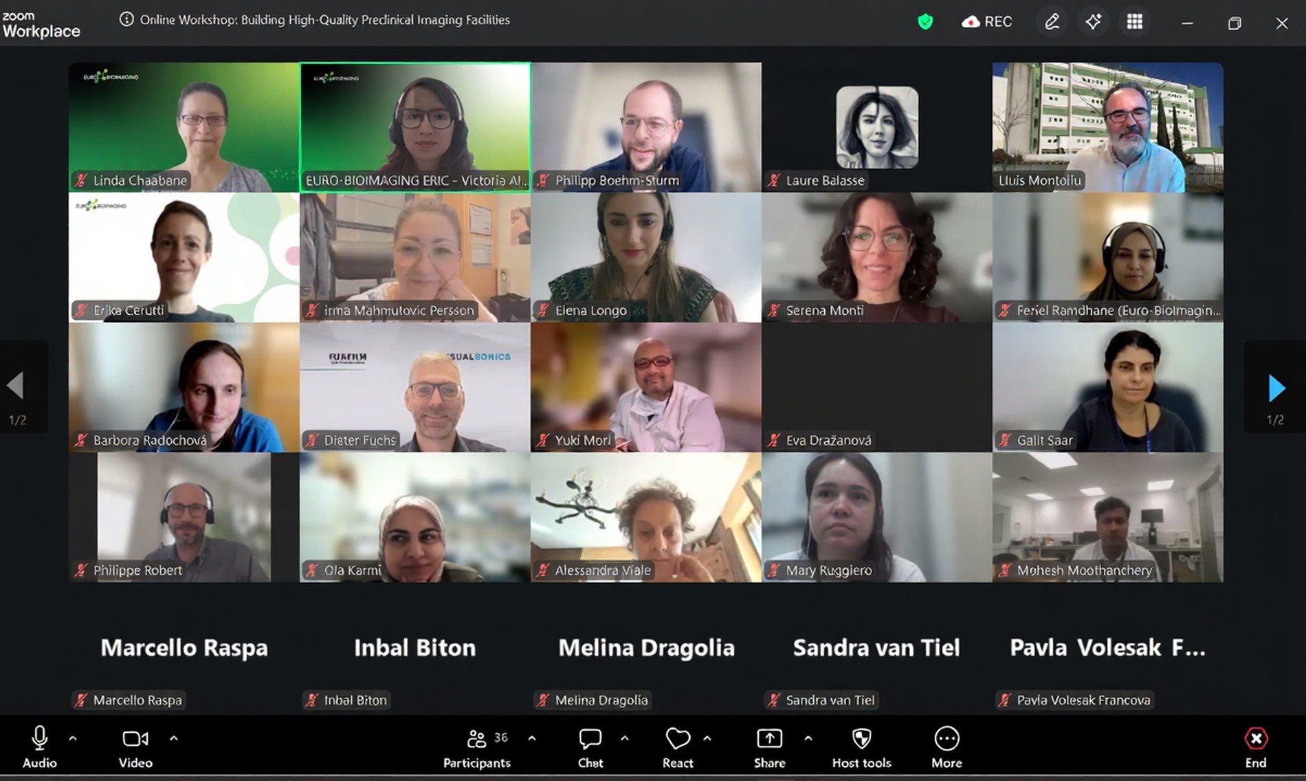
On 2 July 2026, Euro-BioImaging hosted the online EVOLVE workshop “Building High-Quality Preclinical Imaging Facilities”, bringing together approximately 40 imaging facility staff and…