
March 30, 2026
Euro-BioImaging at the EOSC ESFRI meeting in Milan
Euro-BioImaging was delighted to attend the ESFRI/EOSC Policy Workshop on “EOSC and Research Infrastructures: Opportunities and Strategies” in Milan from March 15-16, represented by…

The Asian tiger mosquito Aedes albopictus is an increasingly present species across the globe. With a voracious appetite and aggressive, day-time feeding tactics, female tiger mosquitoes are a nuisance, but also a risk for humans and other animals - as they can transmit a range of diseases such as Zika, chikungunya, and dengue viruses. Irene Arnoldi1, a postdoctoral researcher in the laboratory of Zoology with Professor Paolo Gabrieli at the University of Milan - Italy, has been studying the Asian tiger mosquitoes’ sensory mechanisms to better understand viral transmission and infection. In particular, she has been interested in taking a closer look at the organisation of cells within the labrum, and specifically, the sensory neurons embedded in the labrum sensilla, the mosquitoes' tasting and biting systems. As the structural components of these tiny organs cannot be seen with a normal microscope, she needed to work with high-end volume electron microscopy. Supported by funding from the ISIDORe project, she brought her project to the Euro-BioImaging’s Advanced Light & Electron Microscopy Prague Node, where Frantisek Kitzberger, Jiří Týč and Aleš Benda designed and developed a full pipeline to support her research. Below please find an explanation of how the facility supported this important infectious disease research project.
1Currently working at the Radboud University Medical Center in Nijmegen, The Netherlands
Article written by Frantisek Kitzberger and Aleš Benda
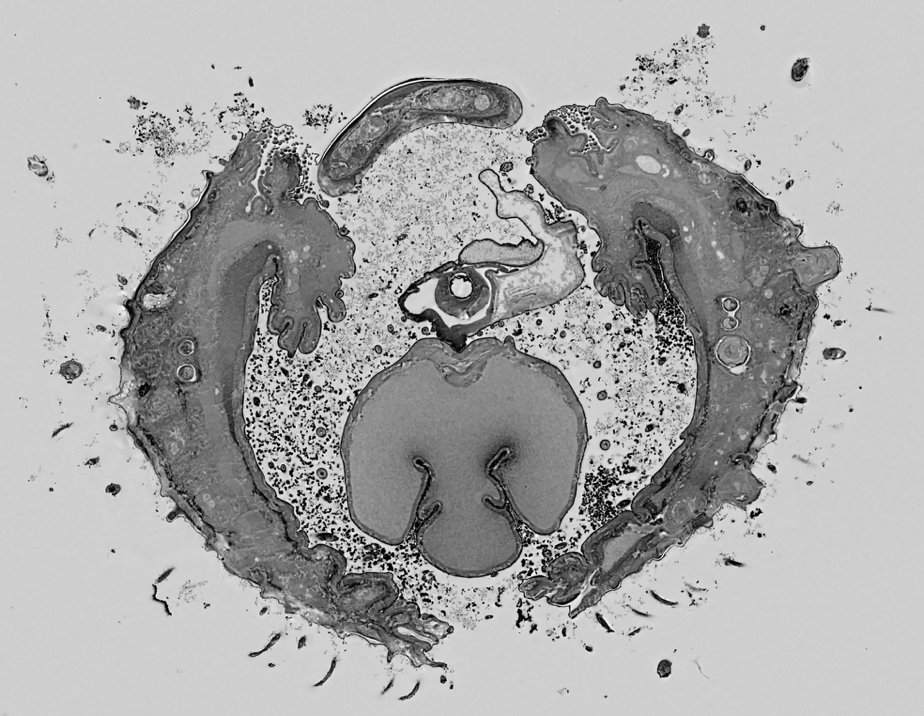
Thanks to support from the ISIDORe-funded access to Euro-BioImaging facilities, and in collaboration with Irene Arnoldi, we were able to successfully carry out a 3D ultrastructural analysis of the proboscis tip of Aedes albopictus, an important vector of arboviruses such as Zika, dengue, and chikungunya. The objective was to clarify the organization and neuronal connectivity of different types of labral sensilla potentially involved in gustatory and mechanosensory perception during blood feeding.
The project goals, as outlined in the original proposal, were fully achieved:
The obtained 3D datasets suggested that the distal labrum tip is connected with the neurons in a slightly different way than was expected, providing information that is otherwise not obtainable by standard 2D imaging.
This project demonstrates how Euro-BioImaging infrastructure facilitated technically challenging volume EM on small and heavily sclerotized arthropod structures. Moreover, the methodological developments and imaging protocols we established may be applied in future to other mosquito species or blood-feeding arthropods.
Access to expert advice, advanced imaging technologies, and structured collaboration with the Prague Euro-BioImaging Node played a key role in the project’s success. The ISIDORe framework enabled us to complete this work without institutional barriers and with strong support throughout the process.
March 30, 2026
Euro-BioImaging was delighted to attend the ESFRI/EOSC Policy Workshop on “EOSC and Research Infrastructures: Opportunities and Strategies” in Milan from March 15-16, represented by…
March 30, 2026
Euro-BioImaging was delighted to attend the Public Awareness & Engagement of Research Infrastructures (PAERI) conference, represented by External Communications Officer, Marianna Childress-Poli. This year’s…

March 26, 2026
The Madrid Advanced Microscopy Center (MAdMiC) is the first Euro-BioImaging Node in Madrid (Spain). It is formed through the collaboration and close work of…