Phantom Limb Pain (PLP), i.e., the pain perceived in an amputated body part, is a very debilitating condition for amputated patients. However, its origin is not fully understood and, for that reason, the available treatment options are limited and not very effective. Carolina Travassos, a PhD student at the University of Coimbra in Portugal, has been working to better understand this condition since 2019, when she started her doctoral research project at the Coimbra Institute for Biomedical Imaging and Translational Research (CIBIT) part of the Institute for Nuclear Sciences Applied to Health (ICNAS), in collaboration with Siemens Healthcare, Portugal. In 2022, her research took her to Euro-BioImaging Dutch High Field Imaging Hub to use an ultra-high field magnetic resonance imaging (MRI) scanner. She explains how this visit supported her research in the interview below.
Carolina Travassos: My PhD project, entitled Unraveling neural circuitries underlying phantom limb pain in the human brain, has been developed at CIBIT in close collaboration with Siemens Healthcare, Portugal. It is focused on Phantom Limb Pain. Since current findings associate limb amputation with significant changes in the sensorimotor network, namely in the primary somatosensory cortex (S1), my project aims to understand the contribution of the S1 to PLP. We hope that a better knowledge of the somatotopic arrangement of S1 will help a better characterization of the neural basis of somatosensory processing and, this way, improve the understanding of the clinical consequences of dysfunctions in the somatosensory cortex, such as PLP. So, the ultimate goal of my project is to help to clarify the underlying mechanisms behind PLP and contribute to the development of more effective therapeutic approaches for this condition. This project wouldn’t be possible without the support and commitment of my supervisors, Professor Miguel Castelo-Branco, Dr. Teresa Sousa, and Dr. Bruno Direito, to whom I am very grateful.
Why did you apply for access with Euro-BioImaging?
Carolina Travassos: The Center for Image Sciences of the University Medical Centre (UMC) at Utrecht offers state-of-the-art neuroimaging facilities, namely a 7T human MRI scanner. This scanner allows the acquisition of reliable functional maps of S1 with submillimeter spatial resolution, enabling the distinction of the somatotopic organization of S1. This was a valuable addition to my work because at CIBIT/ICNAS we just have a 3T MRI scanner.
Besides, the research group with whom I collaborated at UMC (headed by Professor Natalia Petridou) has extensive work on the somatotopy of somatosensory and motor cortices. Actually, they developed a model to characterize the average receptive field properties of neuronal populations in human somatosensory and motor cortices. This model, called Bayesian population receptive field (pRF), was already successfully applied to the cortical representation of the fingers in the S1. In my work, we propose the utilization of this model for the entire upper limb, which is a novelty regarding what has been done before.
Lastly, the UMC facilitates somatosensory stimulators (based on the piezoelectric effect) approved to use in the magnetic resonance environment. This is a very reliable method of somatosensory stimulation and its feasibility to elicit responses in S1 has already been demonstrated.
How has visiting a Euro-BioImaging Node benefitted your project, your science, or your career?
Carolina Travassos: Visiting a Euro-BioImaging Node was an incredibly valuable experience for me both personally and professionally. In terms of my project, we acquired ultra-high-field functional magnetic resonance imaging (fMRI) data using piezoelectric stimulation in the entire arm of healthy participants. The collaboration also included data and methodological sharing (e.g., preprocessing and analysis). For these reasons, having the opportunity to develop this project at UMC was of utmost importance to my research objectives. In general, access to state-of-the-art imaging technologies and equipment that are not available in my home institution allowed me to gather more detailed and precise data for my research. It also allowed me to connect with leading experts in the field, who have provided me with guidance, feedback, and support. This will undoubtedly benefit my science and career in the long term.
In addition, my visit has allowed me to network and build relationships with other researchers, which has given me exposure to diverse perspectives and approaches to scientific research. This has broadened my horizons and inspired me to think more creatively about my work. Overall, I believe that visiting a Euro-BioImaging Node was a great experience that has enriched my research, expanded my knowledge, and opened up the way I see my future.
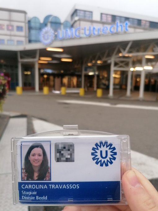
Photo during Carolina's visit to the Dutch High Field Imaging Node
How did you prepare for this experience?
Carolina Travassos: Since it was a very short stay, my supervisors and I met with the team from UMC (Professor Natalia and Dr. Wouter Schellekens) beforehand to discuss and prepare the experiment. Besides, all the necessary paperwork for my stay and entrance at UMC was also prepared in advance. This allowed us to start acquiring MRI data on my second day at UMC and process/analyze that data on the remaining days.
How is this different from what you do at your home institution?
Carolina Travassos: In fact, it was not that different from what I am accustomed to during my workday at CIBIT/ICNAS. I’m used to working in the magnetic resonance environment (which is a highly controlled environment due to a strong magnetic field) and acquiring/analyzing fMRI data. The main difference was the quality of the acquired data. 7T scanners provide images with a higher signal-to-noise ratio, spatial resolution, and image quality in general. On the other hand, there are also some challenges associated with 7T data, such as increased susceptibility artifacts and greater image distortion. These issues require careful management and optimization of imaging protocols (to ensure the best possible image quality), as well as optimized pre-processing pipelines. Both aspects were covered during my stay at UMC, making it a valuable learning experience for me.
Article written by: Carolina Travassos
More news from Euro-BioImaging
July 28, 2026
Global BioImaging Advance the Conversation on Sustainable Biomedical Imaging Facilities
How can biomedical imaging facilities remain scientifically excellent while becoming more financially, environmentally, and operationally sustainable? These questions were at the heart of…
Moara Lemos: New insights from Global BioImaging’s Job Shadowing programme
Moara Lemos is a Brazilian researcher and imaging scientist. Her award winning research pushes the boundaries of structural biology, using advanced cryo-electron microscopy…
A Multiplexed Vision: Highlights from the Special Edition Virtual Pub on Spatial Transcriptomics
On June 12th, Euro-BioImaging hosted its latest Special Edition Virtual Pub, bringing together over 200 researchers and technology developers working at the cutting…